
Καρκίνος του τραχήλου της μήτρας: επιδημιολογία, αιτιολογία, παθογένεση και κύριοι ιστολογικοί τύποι.
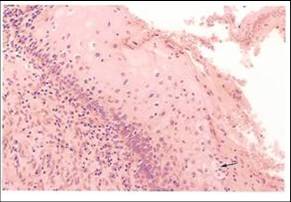
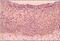
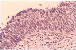
Ιστολογικά χαρακτηριστικά του CIN
- Δομικές αλλαγές στο επιθήλιο:
- Αντικατάσταση ολοκλήρου του πάχους του πλακώδους επιθηλίου του τραχήλου της μήτρας από μη φυσιολογικό επιθήλιο.
- Αδιαφοροποίητα άτυπα κύτταρα με υψηλή πυρηνο /πλασματική αναλογία αντικαθιστούν την βασική στιβάδα με απώλεια προσανατολισμού. Το επίπεδο στο οποίο βρίσκονται αυτά τα αδιαφοροποίητα κύτταρα προσδιορίζει τον βαθμό του CIN.
- Άτυπη ωρίμανση και άτυπη αρχιτεκτονική διαμόρφωση του επιθηλίου.
- Αυξημένη κυτταροβρίθεια του επιθηλίου.
- Κυτταρολογικές αλλαγές του επιθηλίου
- Παρουσία μιτώσεων – συχνά άτυπων
- Πλειομορφία στους πυρήνες, ποικιλία στο σχήμα και το μέγεθος
- Υπερχρωμασία
 |
 |
 |
| CIN1 : note undifferentiated abnormal cells with high n/c ratio occupying the lower 1/3 of the epithelium and abnormal maturation with loss of stratification in the upper 2/3 of the epithelium. | CIN2: note abnormal cells with high n/c ratio occupying 2/3 of the epithelium,showing loss of polarity and presence of mitotic figure .Some abnormal maturation is seen in upper 1/3 of epithelium | CIN3: note abnormal cells with high n/c ratio occupy full thickness of epithelium. They show loss of polarity, pleomorphism, hyperchromasia and mitotic figures |
Η Τραχηλική Ενδοεπιθηλιακή Νεοπλασία μπορεί να εξελιχθεί σε διηθητικό καρκίνο εάν δεν θεραπευτεί.
- Ο κίνδυνος της εξέλιξης αυξάνεται με τον βαθμό του CIN
- Υψηλότερος κίνδυνος εξέλιξης στο CIN3, χαμηλότερος στο CIN1
- Μπορεί να παραμείνει αμετάβλητο
- Για την εξέλιξη μπορεί να χρειαστούν έως 20 χρόνια
- Το CIN1 μπορεί να υποχωρήσει
- Δεν υπάρχουν αυστηρά όρια μεταξύ του CIN1 /2 /3
Αποδείξεις που στηρίζουν την άποψη ότι το CIN που δεν υποβάλλεται σε θεραπεία μπορεί να οδηγήσει σε διηθητικό καρκίνο
- Έμμεσες αποδείξεις ότι το CIN1, 2, και 3 είναι συνεχόμενες αλλοιώσεις (lesional continuum) βασίζονται σε πειραματικές μελέτες. Άμεσες αποδείξεις ότι το CIN είναι πρόδρομος του διηθητικού καρκινώματος βασίζονται σε προοπτικές μελέτες γυναικών που δεν υποβλήθηκαν σε θεραπεία του CIN. Ακολουθεί μία σύντομη ανασκόπηση αυτών των μελετών.
Πειραματικές μελέτες οι οποίες υποστηρίζουν ότι το CIN1, 2 και 3 είναι συνεχόμενες αλλοιώσεις (lesional continuum)
- Αυτοραδιογραφικές μελέτες της δραστηριότητας πολλαπλασιασμού του φυσιολογικού επιθηλίου του τραχήλου, δυσπλασία και καρκίνωμα in situ. Αυτές διεξήχθησαν από τον Richart (1963) ο οποίος χρησιμοποίησε ως σήμανση την τριτιωμένη θυμιδίνη σε κύτταρα που συνέθεταν DNA σε βιοψίες τραχήλου. Διαπίστωσε ότι ο χρόνος της αναγέννησης του επιθηλίου ελαττώνεται με την σοβαρότητα της αλλοίωσης του τραχήλου και ήταν μικρότερος στο καρκίνωμα in situ.
- DNA κυτταροφωτομετρικές μελέτες από βιοψίες φυσιολογικού και δυσπλαστικού επιθηλίου και καρκινώματος in situ. Ο Willbanks et al (1967) μέτρησε το περιεχόμενο του DNA των κυττάρων σε τομές χρησιμοποιώντας χρώση Feulgen, και μικροσπεκτροφωτομετρική μέθοδο και έδειξε ότι η ποσότητα του DNA αυξάνεται όσο αυξάνεται η σοβαρότητα της τραχηλικής αλλοίωσης και ο βαθμός της απόκλισης της ποσότητας του DNA από το φυσιολογικό είναι μεγαλύτερος για το καρκίνωμα in situ.
- Μελέτες καρκινογένεσης στο επιθήλιο του τραχήλου της μήτρας σε ζώα. Σε αυτές τις μελέτες οι τράχηλοι μυών βαφόταν με καρκινογόνο ουσία (3,4 – βενζοπυρένιο) δύο φορές την εβδομάδα [Rubio και Lagerhof (1974)]. Βρήκαν ότι αλλαγές του τύπου της ήπιας δυσπλασίας αναπτύχθηκαν στο τραχηλικό επιθήλιο των μυών μετά από δύο μήνες, μέτρια δυσπλασία μετά από τέσσερις μήνες και σοβαρή δυσπλασία, καρκίνωμα in situ και μικροδιηθητικός καρκίνος μετά από πέντε μήνες.
- Άλλες ενδιαφέρουσες πειραματικές εργασίες, που έγιναν, συμπεριλαμβάνουν κυτταρογενετικές μελέτες (Springs et al 1971), υπερμικροσκοπικές μελέτες (Murphy et al 1975) και μελέτες κυτταροκαλλιέργειας καρκινικών κυττάρων (Richart 1964).
- Προοπτικές μελέτες σε γυναίκες με CIN
- Αποδείξεις που ενισχύουν την άποψη ότι το CIN είναι πρόδρομος του διηθητικού καρκίνου έχουν αποκτηθεί από προοπτικές μελέτες σε γυναίκες με δυσπλασία οι οποίες δεν θεραπεύτηκαν, αλλά ήταν κάτω από κυτταρολογική και κολποσκοπική επίβλεψη ανά ποικίλα περιοδικά χρονικά διαστήματα. Αν και οι μελέτες θεωρούνται σήμερα από ηθικής άποψης ανεπίτρεπτες, μας παρέχουν σαφείς αποδείξεις ότι η δυσπλασία εάν δεν θεραπευτεί μπορεί να εξελιχθεί σε καρκίνωμα in situ και διηθητικό καρκίνο μετά από χρόνια. Έχουν επίσης αποδείξει ότι μερικές δυσπλασίες υποχωρούν. Αρκετές μελέτες συμπεριέλαβαν μικρό αριθμό γυναικών οι οποίες παρακολουθήθηκαν ιατρικώς για μικρό χρονικό διάστημα και τα συμπεράσματα στα οποία κατέληξαν δεν είναι ασφαλή.
- Ευλόγως, η πιο πειστική έρευνα είναι αυτή που διεξήχθη από τον Mc Indoe et al από την Νέα Ζηλανδία, όπου γυναίκες με καρκίνωμα in situ παρακολουθήθηκαν κυτταρολογικά, υποθέτοντας ότι ο κίνδυνος της εξέλιξης ήταν πολύ χαμηλός (Green και Donovan 1970, Green 1974). Προοπτικές μελέτες έδειξαν ότι ο κίνδυνος του διηθητικού καρκίνου, αφού έχει προηγηθεί διάγνωση καρκινώματος in situ, ήταν πολύ υψηλός σε γυναίκες οι οποίες αντιμετωπίστηκαν με αυτόν τον τρόπο. Δέκα οχτώ τοις εκατό (18%) των γυναικών ανέπτυξαν διηθητικό καρκίνο μετά από 10 χρόνια και 36% μετά από 20 χρόνια.
- Οι μελέτες που περιγράφτηκαν παραπάνω επιδεικνύουν πειστικά ότι εάν δεν θεραπευτούν οι CIN αλλοιώσεις μπορούν να εξελιχθούν σε διηθητικό καρκίνο. Εντούτοις, δείχνουν επίσης ότι κάποιες CIN αλλοιώσεις μπορούν να υποχωρήσουν. Αυτή η παρατήρηση έχει επιπτώσεις στην αντιμετώπιση των γυναικών με CIN. Επειδή στις ερευνητικές εργασίες που προαναφέρθηκαν, ο βαθμός διαφοροποίησης των αλλοιώσεων σε συγκεκριμένο χρονική στιγμή δεν είναι γνωστός, είναι δύσκολο να είμαστε απολύτως βέβαιοι σε ποιο ακριβώς στάδιο των αλλοιώσεων η πιθανότητα της υποχώρησης είναι μεγαλύτερη από τον κίνδυνο της εξέλιξης της αλλοίωσης. Οι κλινικοί γιατροί τείνουν να συμφωνούν ότι η πιθανότητα υποχώρησης της ήπιας δυσπλασίας (CIN1) είναι υψηλή (30%). Για αυτό, όταν διαγιγνώσκεται κυτταρολογικά CIN1, είναι έτοιμοι να υιοθετήσουν την τακτική της αναμονής (τουλάχιστον για 6 μήνες) πριν να αποφασίσουν θεραπεία,.
Author |
Study Group |
Outcome |
Fox 1967 |
278 women with mild dysplasia followed for 11 years without biopsy |
Lesion regressed to normal in 31% , |
Richart & Barron |
557 women with dysplasia |
Rate of progression of all degrees of dysplasia to carcinoma in situ was 80% |
Kinlen and Spriggs 1978
|
Traced 52 British women with abnormal smears (Pap class IV or V) who did not attend for treatment for at least 5.2 years after the abnormal smear report |
Repeat smear was normal in 37% |
Nasiell et al (1986) |
555 women with mild dysplasia followed without biopsy for 12 years |
Lesion regressed in 62% |
Mc Indoe et al (1984) |
948 women with biopsy proven carcinoma in situ followed by cytology only from 5 to 28 years |
22% of 131 women who continued to have abnormal cytology developed invasive cancer |