

Cervical Cytology
Inflammatory changes, specific infections reaction, repair and iatrogenic changes.
| Cause and effect of injury to cervix |
| Non specific inflammatory changes |
| Specific infections |
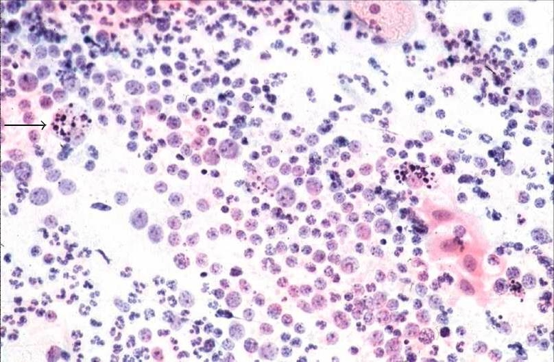
| chronic cervicitis and folicular cervicitis |
| Regeneration and repair |
| Clinical notes |
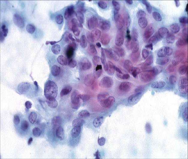
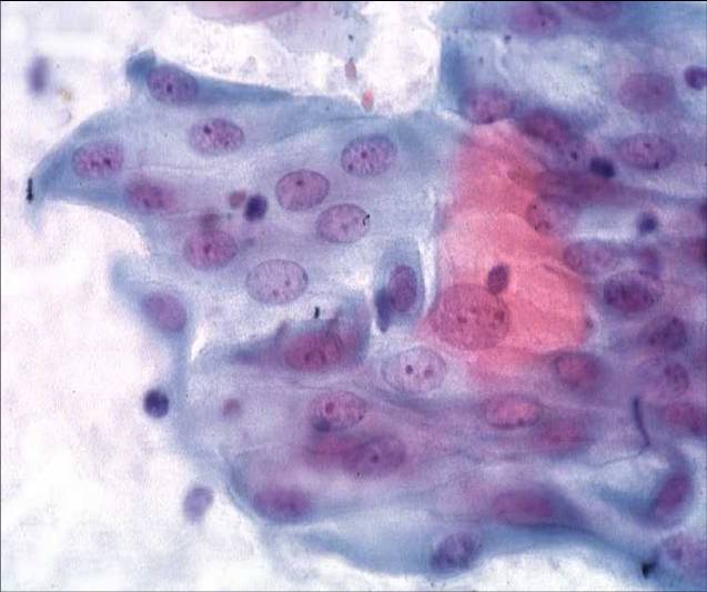
Regeneration and repair
- Healing of the ulcerated or eroded epithelium is effected by proliferation of the basal cell layer of adjacent epithelium and expansion of the reserve cells of the local gland crypts. Initially the denuded area is covered by immature metaplastic cells which eventually transforms into mature squamous or columnar epithelium.
- The degenerative changes associated with inflammation and the regenerative changes associated with repair are often seen in the same smear.
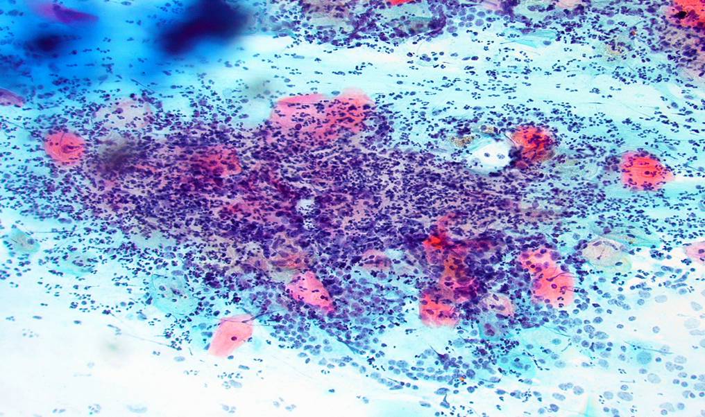
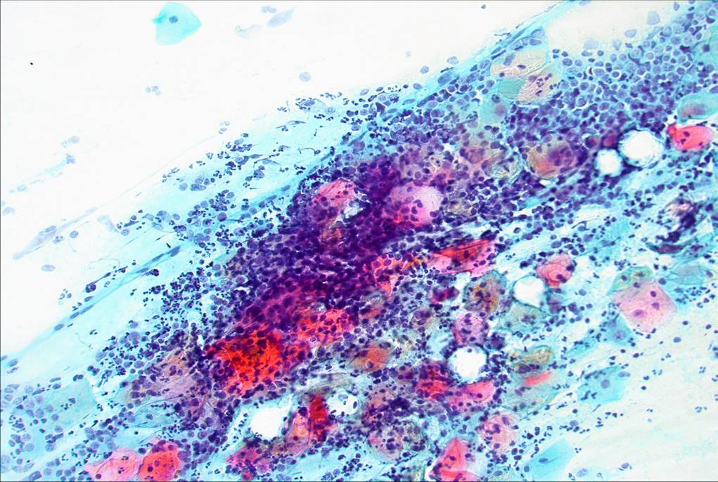
Chronic cervicitis
When injury persists discrete lymphocytes and plasma cells migrate into the inflamed area and can be found in moderate numbers among the polymorphs in the smear. In long standing cervicitis such as that associated with the insertion of a vaginal pessary for vaginal prolapse in a post menopausal woman, the epithelium may show reactive changes such as parakeratosis and hyperkeratosis. These changes may also be seen in the smear.