Cervical Cytology
Inflammatory changes, specific infections reaction, repair and iatrogenic changes.
Cause and effect of injury to cervix
Non specific inflammatory changes
Specific infections
chronic cervicitis and folicular cervicitis
Regeneration and repair
Clinical notes


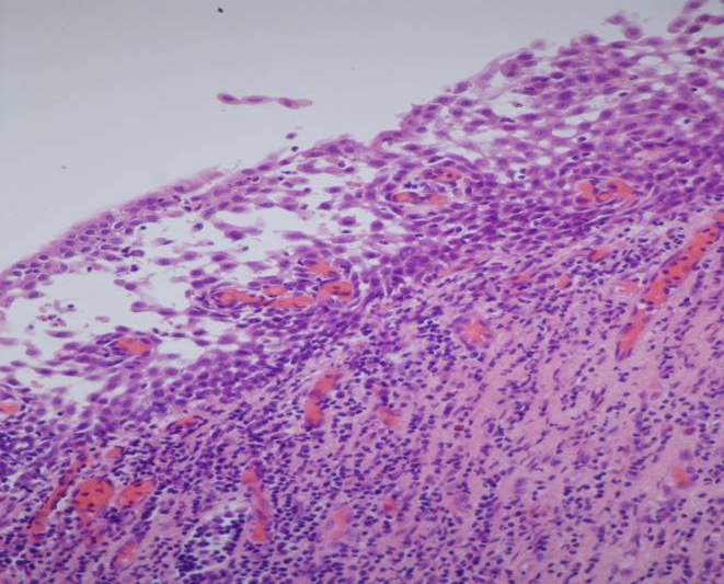
Inflammatory changes in the cervix

Histological section showing inflammatory changes characteristic of acute non specific cervicitis (Click to see text)

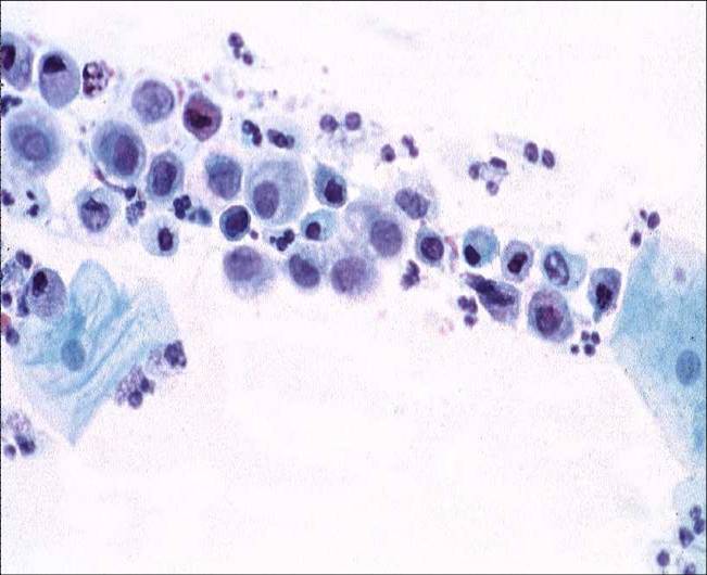
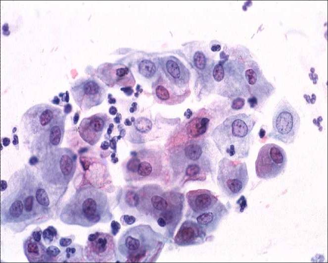
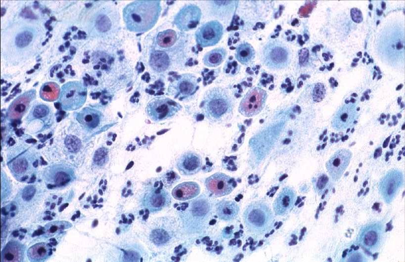
Cytology of  acute non specific cervicitis

The characteristic  features of the smear are:

  1. Epithelial cells which show marked degenerative changes of the cytoplasm and nucleus. The cytoplasm appears frayed  or vacuolated and may exhibit polychromatic staining. It may shrink slightly to produce a perinuclear halo .Occasionally polymorphs are engulfed in the cytoplasm The nuclear changes vary with the severity of the injury. In the earliest stage they show coagulative necrosis with clumping of the chromatin,apparent hyperchromasia and irregularity of the nuclear membrane.   When complete cell lysis supervenes the nuclei become swollen and hypochromatic  and the nuclear structure and membrane become indistinct. Eventually the nucleus shrinks and become pyknotic and karyorrhexis supervenes
  2. Numerous polymorphs embedded in a protein rich exudate. Occasionally the polymorphs are so numerous that they obscure the epithelial cells in the smear and render the smear inadequate for evaluation.
  3. Red blood cells, chains of coccoid bacteria  or bacilli, mucin  and necrotic cell debris may  also be seen in the background.
Non specific cervicitis : Note degenerative changes in the immature metaplastic cells in the inflammatory smear. The epithelial cells show condensation and clumping of the chromatin  in some nuclei and clearing of chromatin in others . The nuclei appear to vary in size . Note vacuolation of the cytoplasm


Cytology of atrophic cervicitis
Atrophic cervicitis : Atrophic cervices are particularly susceptible to infection The smears contain numerous polymorphs and mucus strands and the parabasal Cells show degenerative changes such as nuclear pyknosis and karyorrhexis. Note polychromatic staining of the cytoplasm
Back
Next