Non-gynaecological Cytology
Thyroid cytology
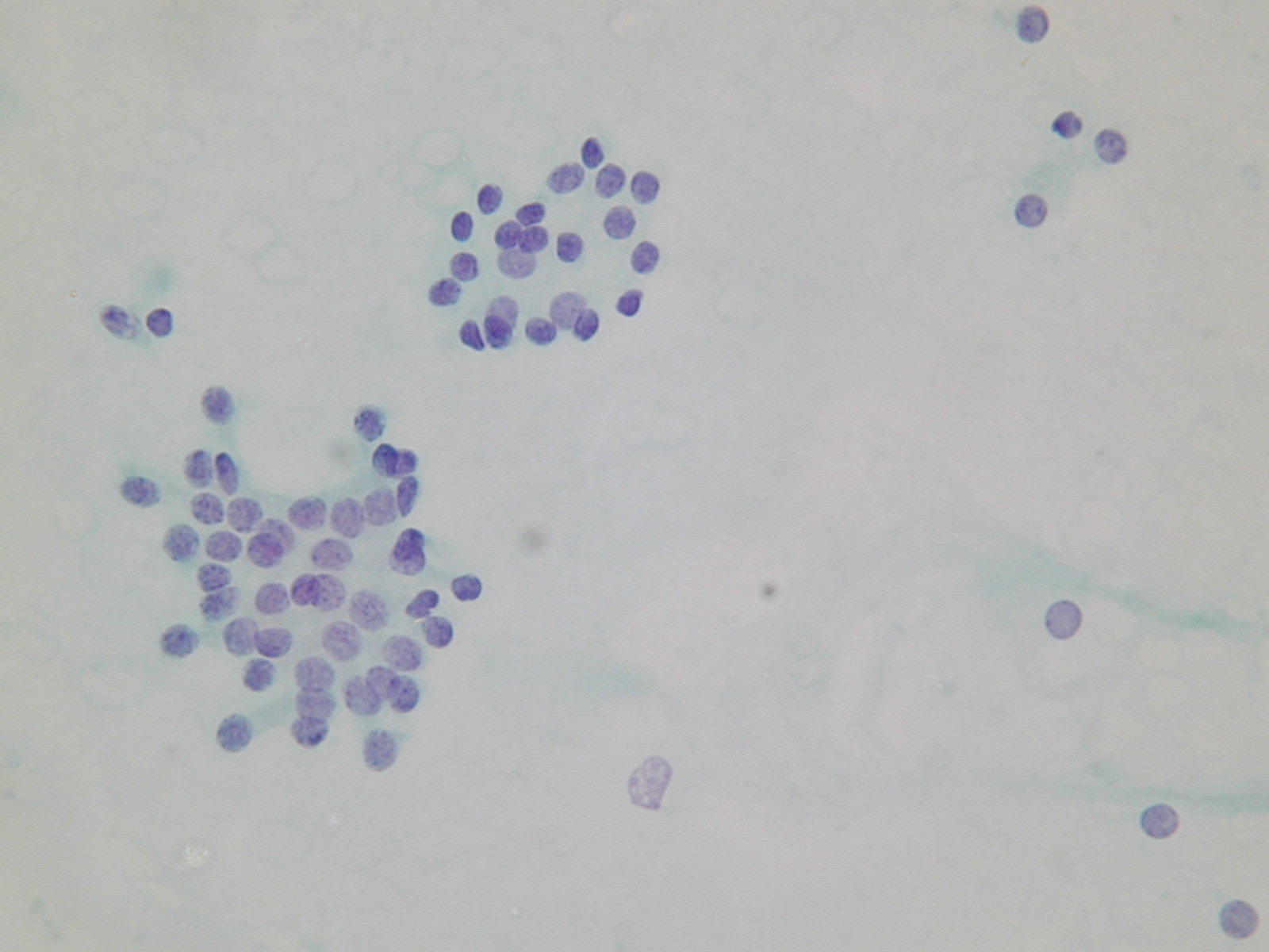
cytologically benign follicular lesions
Cytologic diagnostic features
- low or moderate cellularity
- cohesive cells
- predominantly microfollicular pattern
- uniform, evenly spaced follicular cells
- round nucleus, finely granular chromatin
- scanty or moderate cytoplasm
- few macrophages
- bare nuclei
Colloid is usually abundant, appearing as amorphous blobs or as a thin translucent film with bubbles and linear cracks.
Some benign follicular lesions are hypercellular and even focal cytologic atypia may occur. Occasional large, spindle-shaped cells may be present, representing either reactive stromal elements or altered follicular cells lining areas of cystic degeneration. Focal Hürthle cell change may also occur. If microfollicles are few and atypia is focal, a diagnosis of cytologically benign nodule should be made, even if the specimen is cellular. Patients with this diagnosis have to be followed up at an appropriate interval.
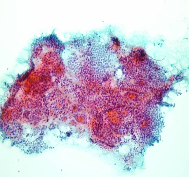
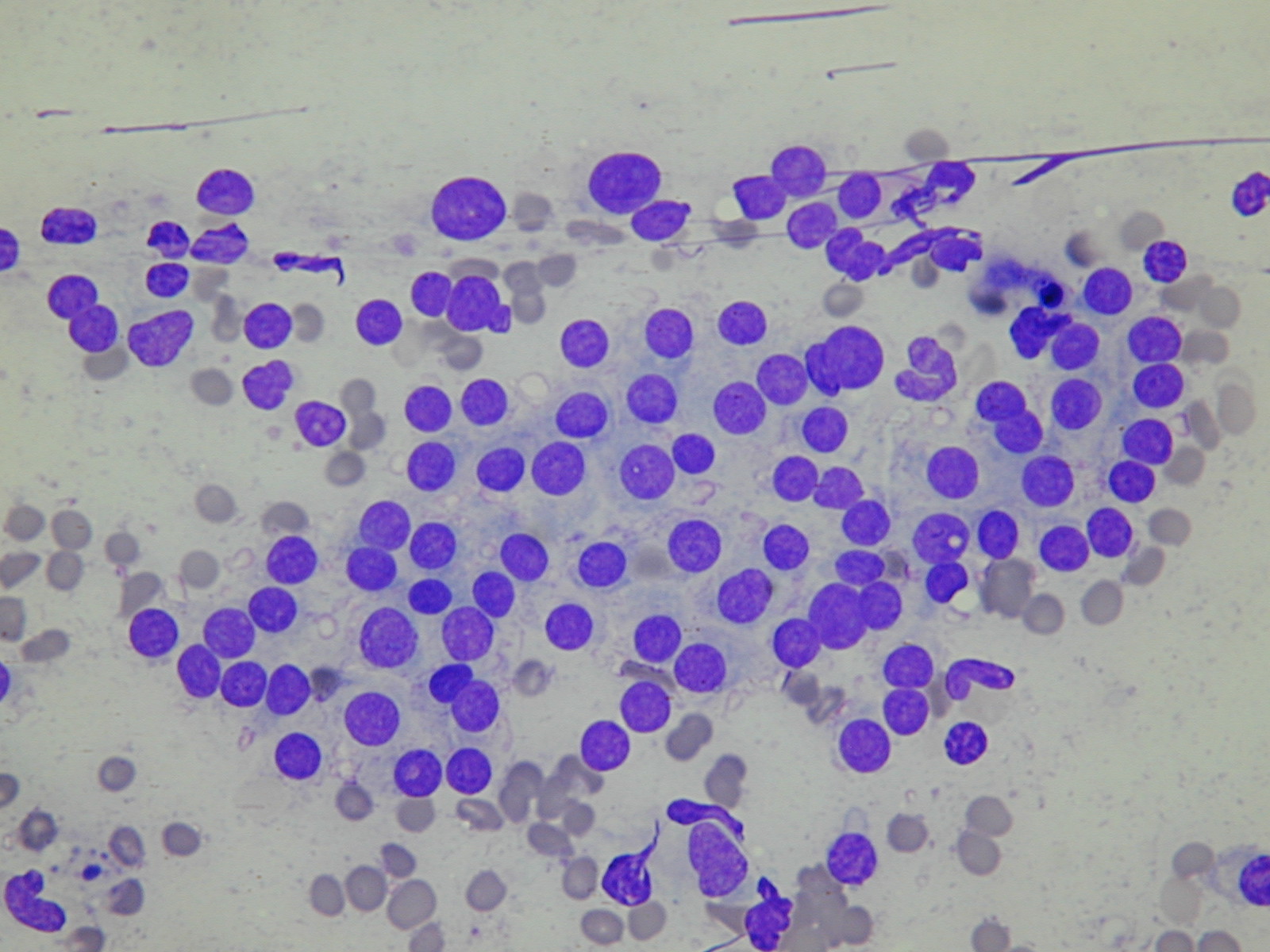
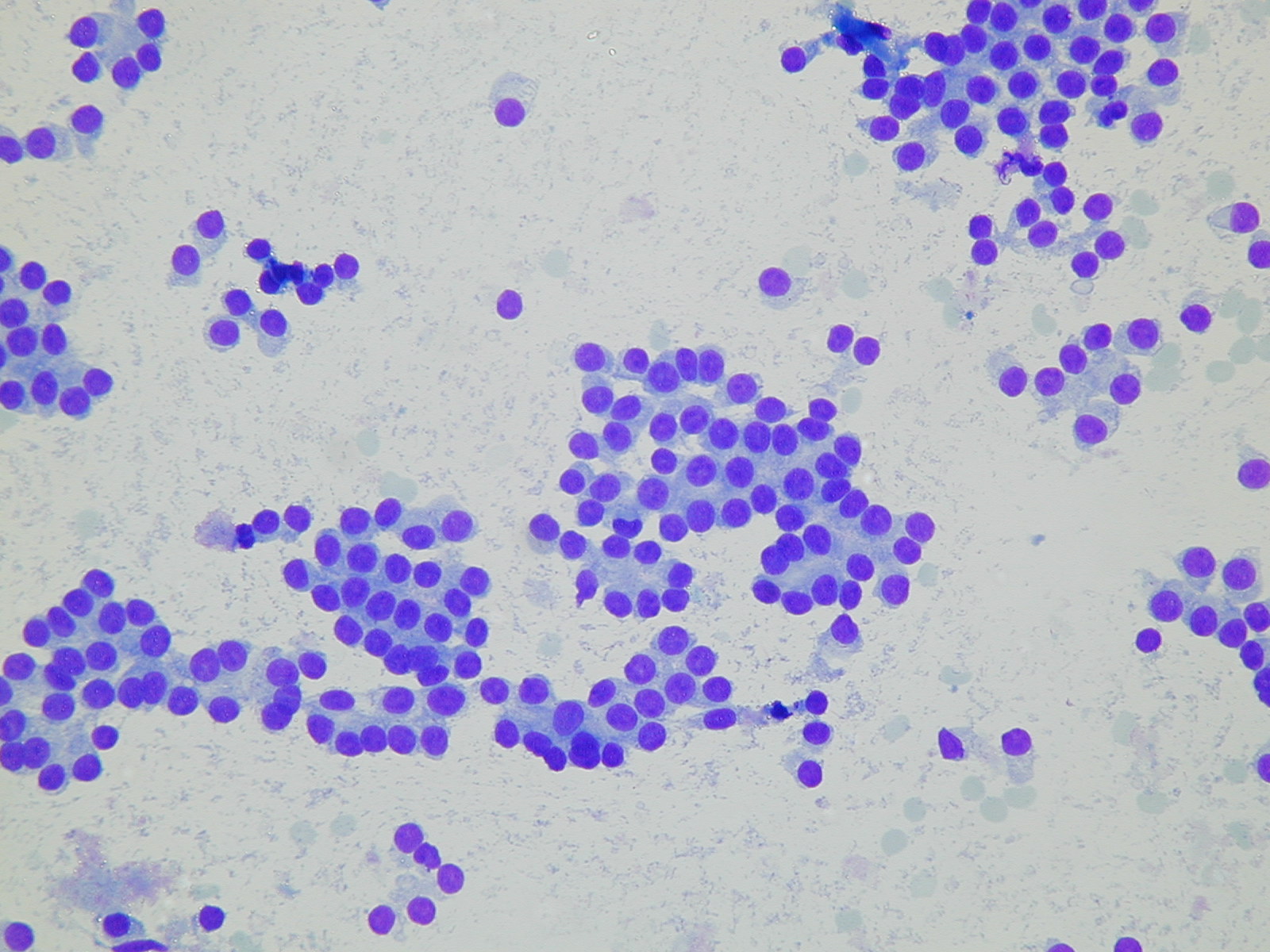
A microbiopsy from a nodular goitre. Bland thyrocytes in a microfollicular arrangement. Bare nuclei and microfollicles.
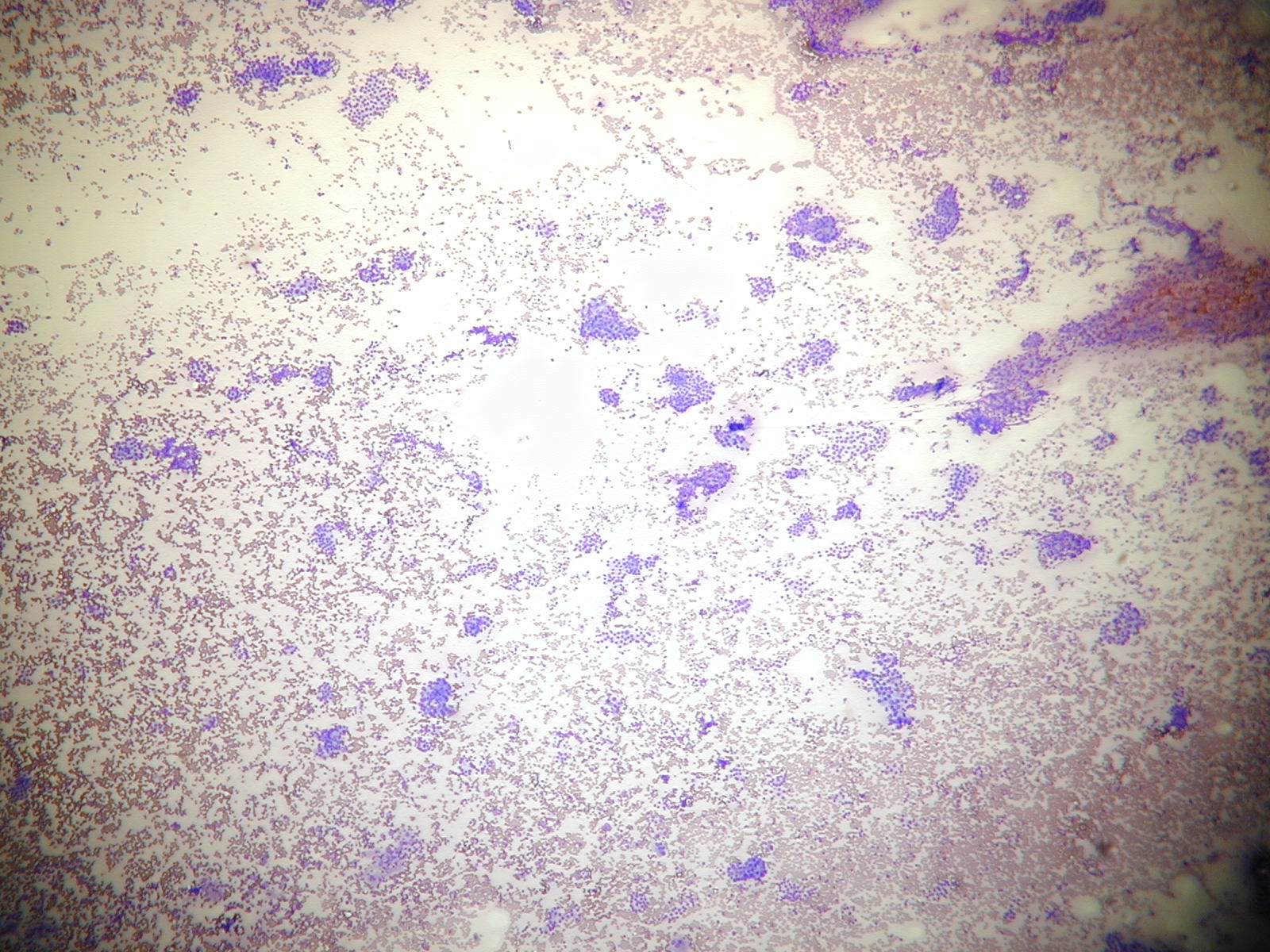
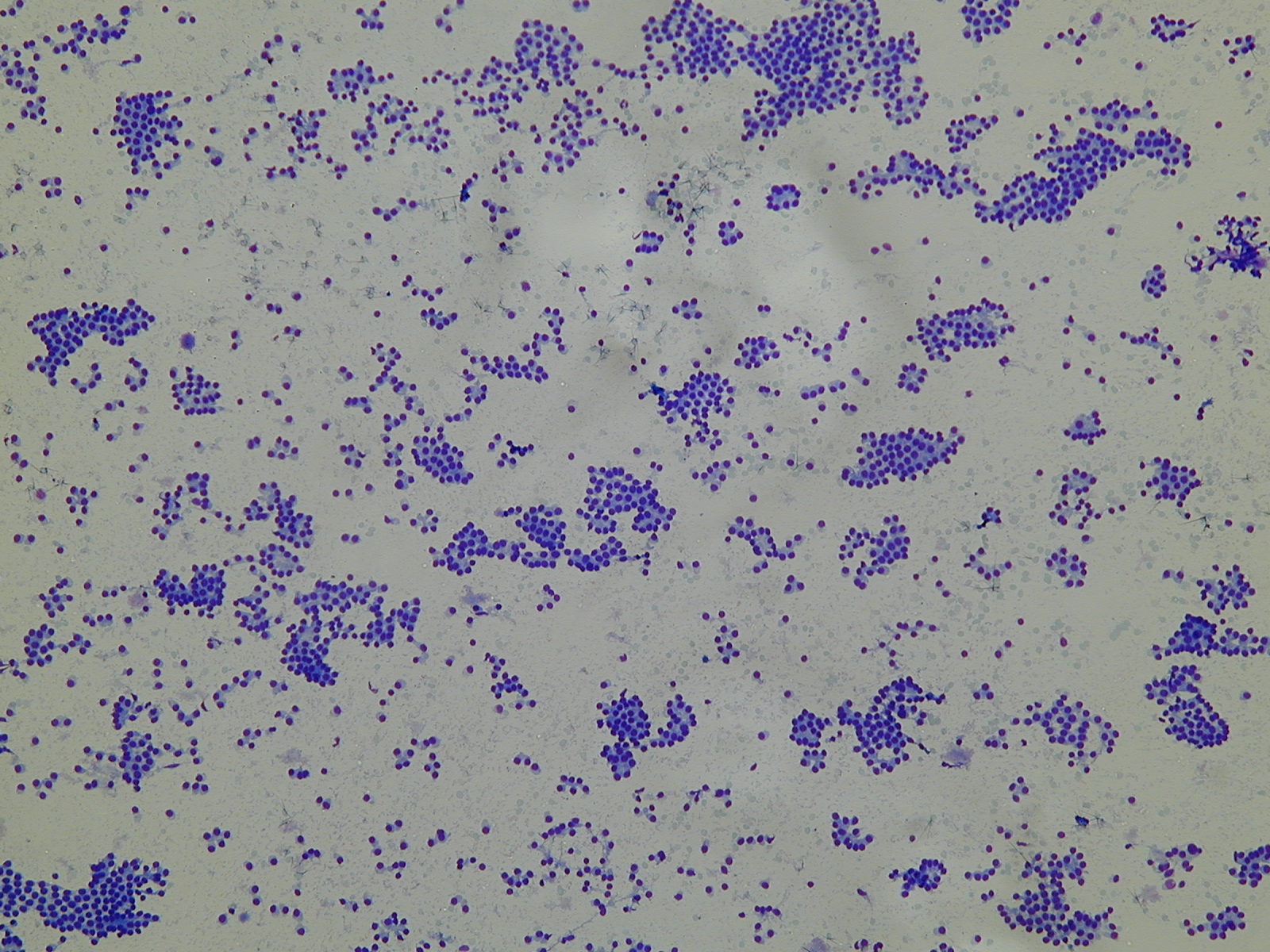
A very low power view of a cellular lesion. At high power the cells are uniform. A trabecular arrangement.
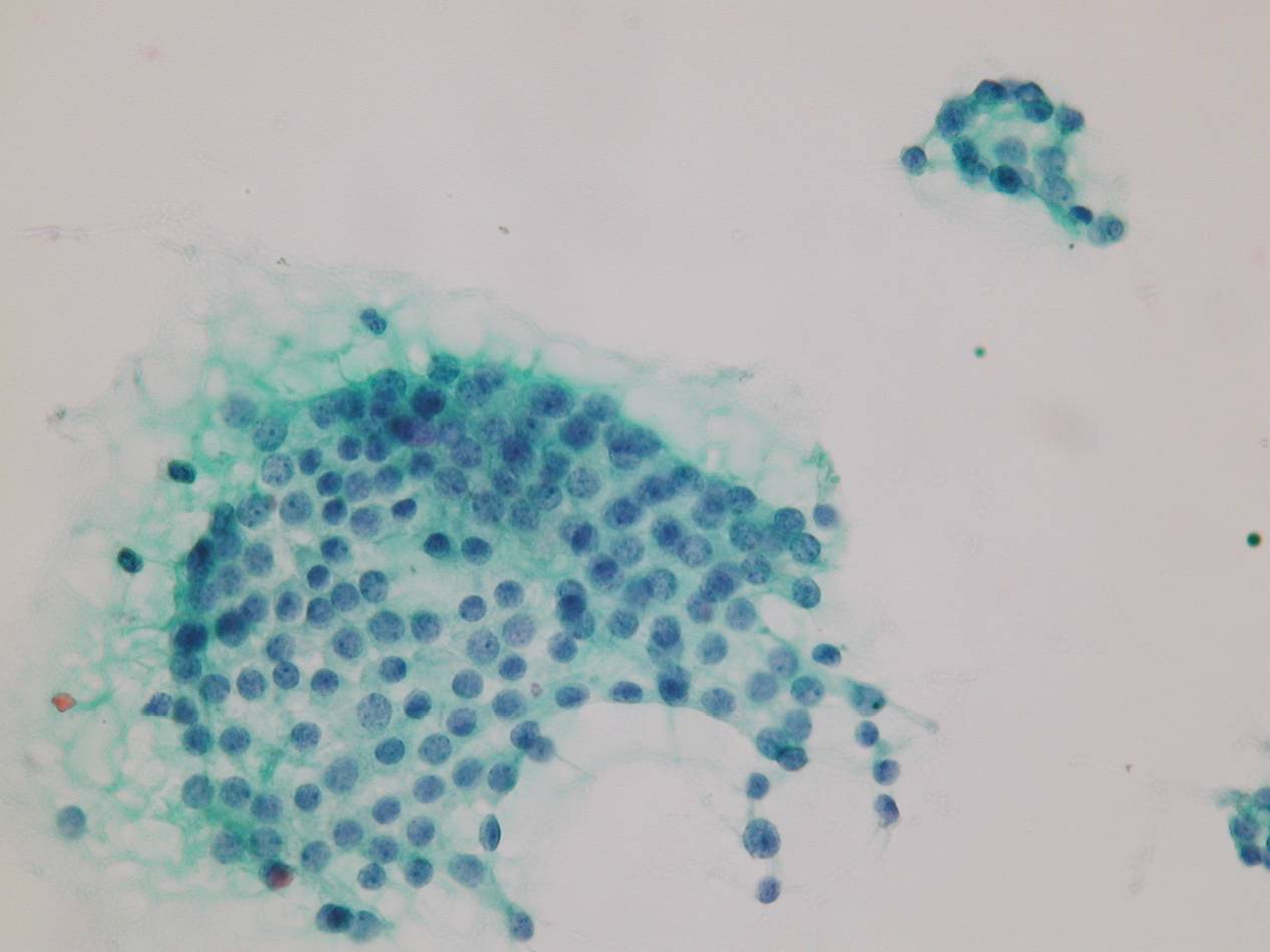
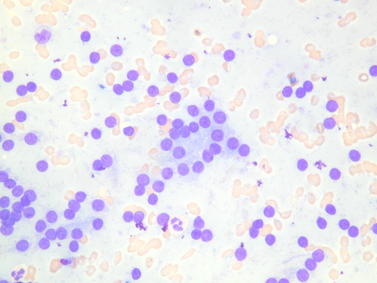
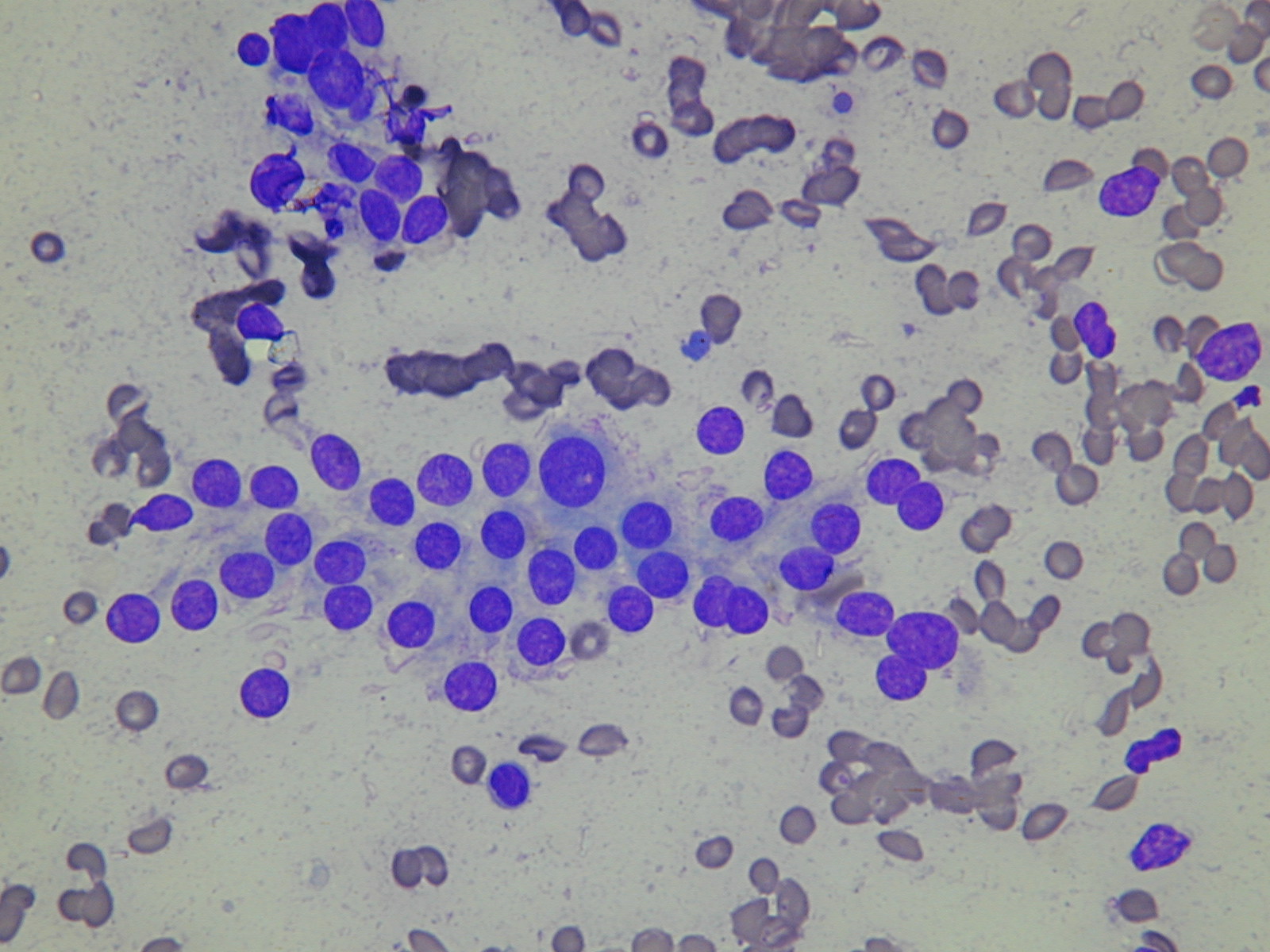
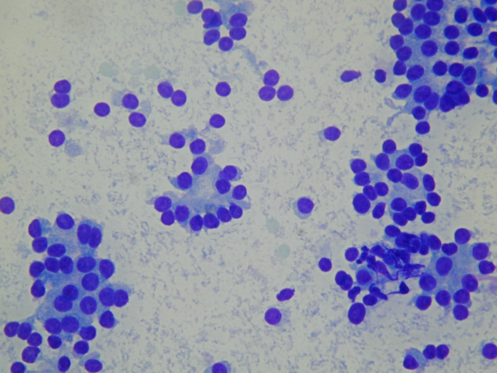
Bland, less cohesive thyrocytes. A cellular aspirate at low power. The cells are uniform and arranged in follicles.
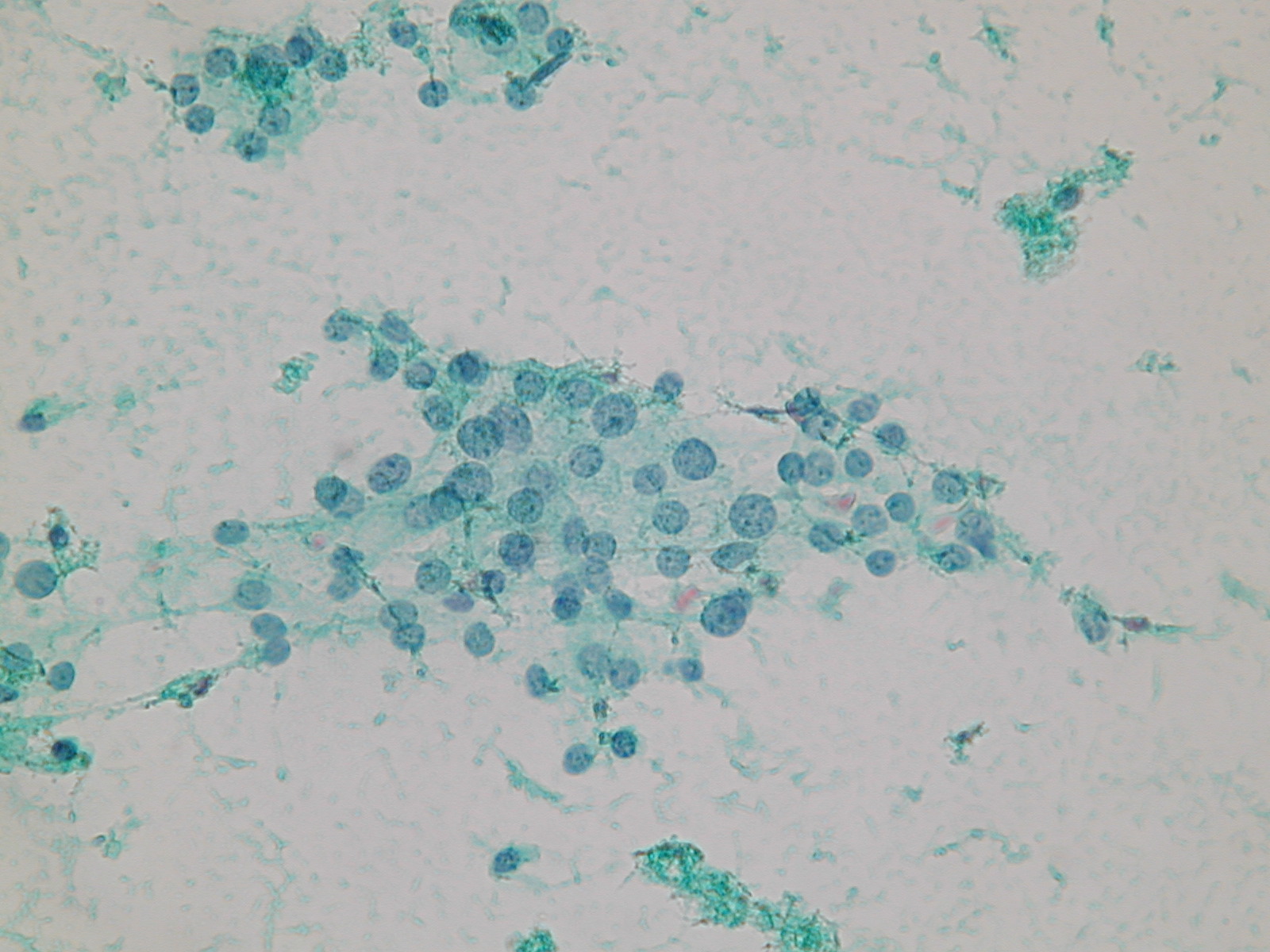
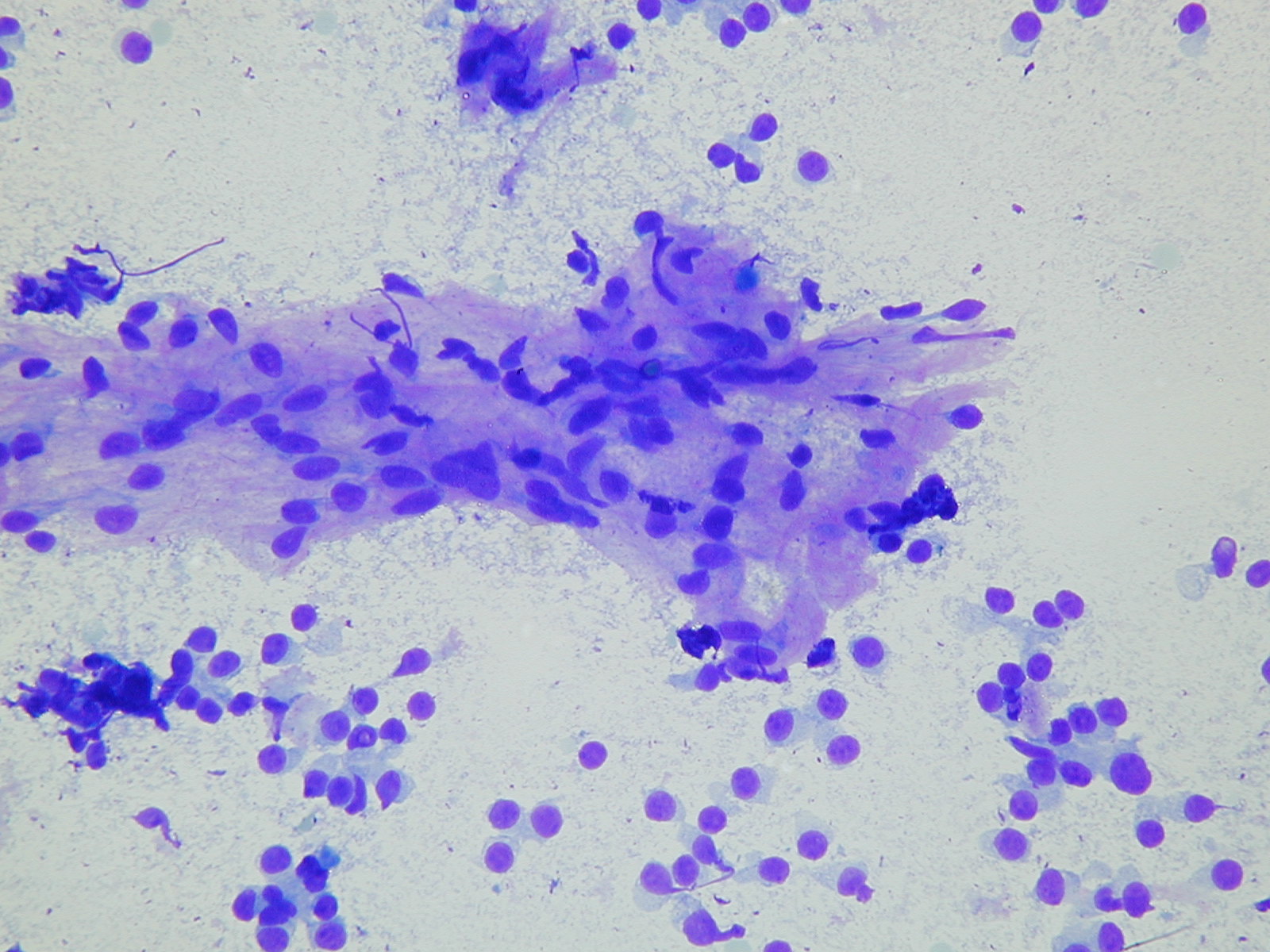
A microfollicular pattern. A stromal fragment with mesenchymal nuclei.
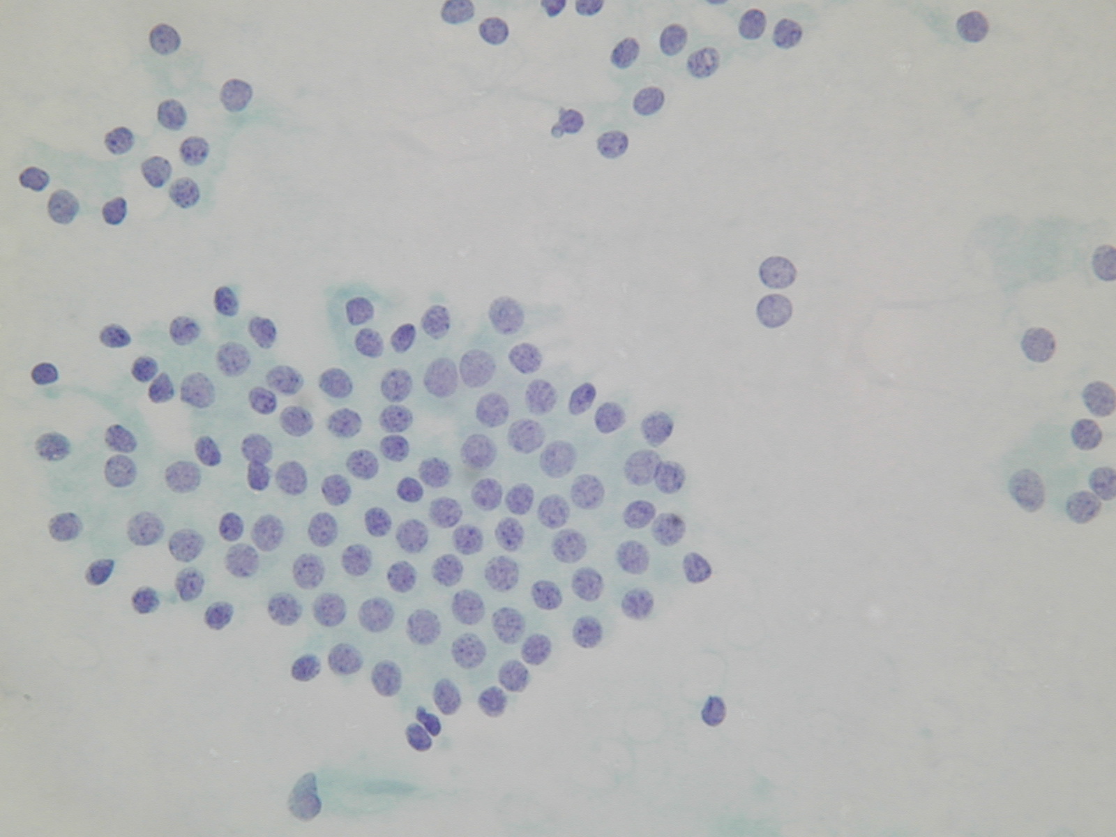
A monolayered sheet of bland thyrocytes. A microfollicular arrangement.