Evaluation of the specimen
Several features will be considered in the evaluation of the specimen:
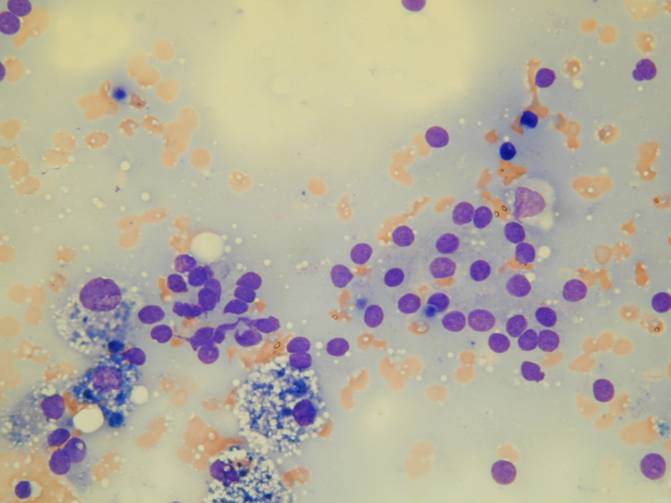
•the type of cells (such as thyrocytes, macrophages, lymphocytes)
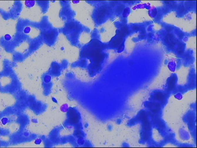
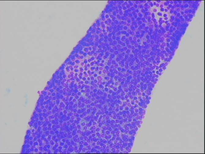
•amount and type of colloid (scanty-abundant, fluid-dense)
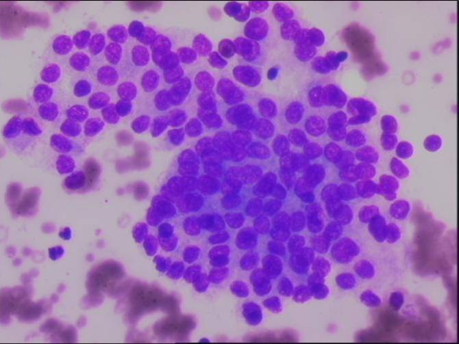
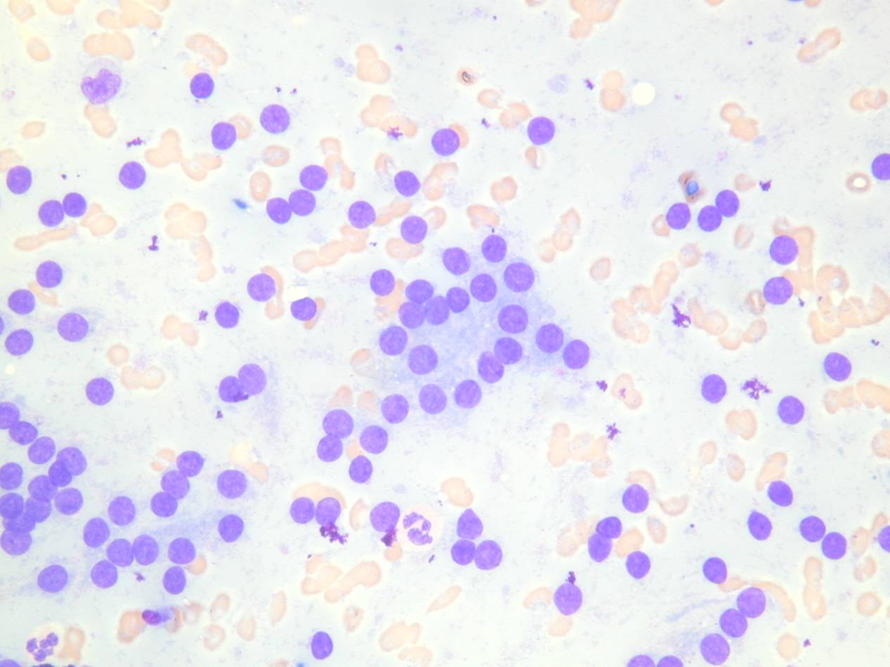
•cellularity (scanty, moderate, marked)
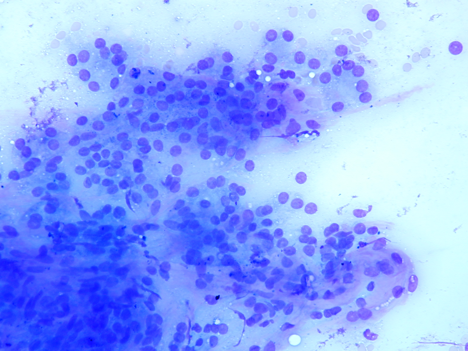
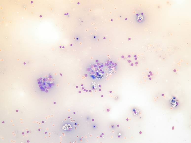
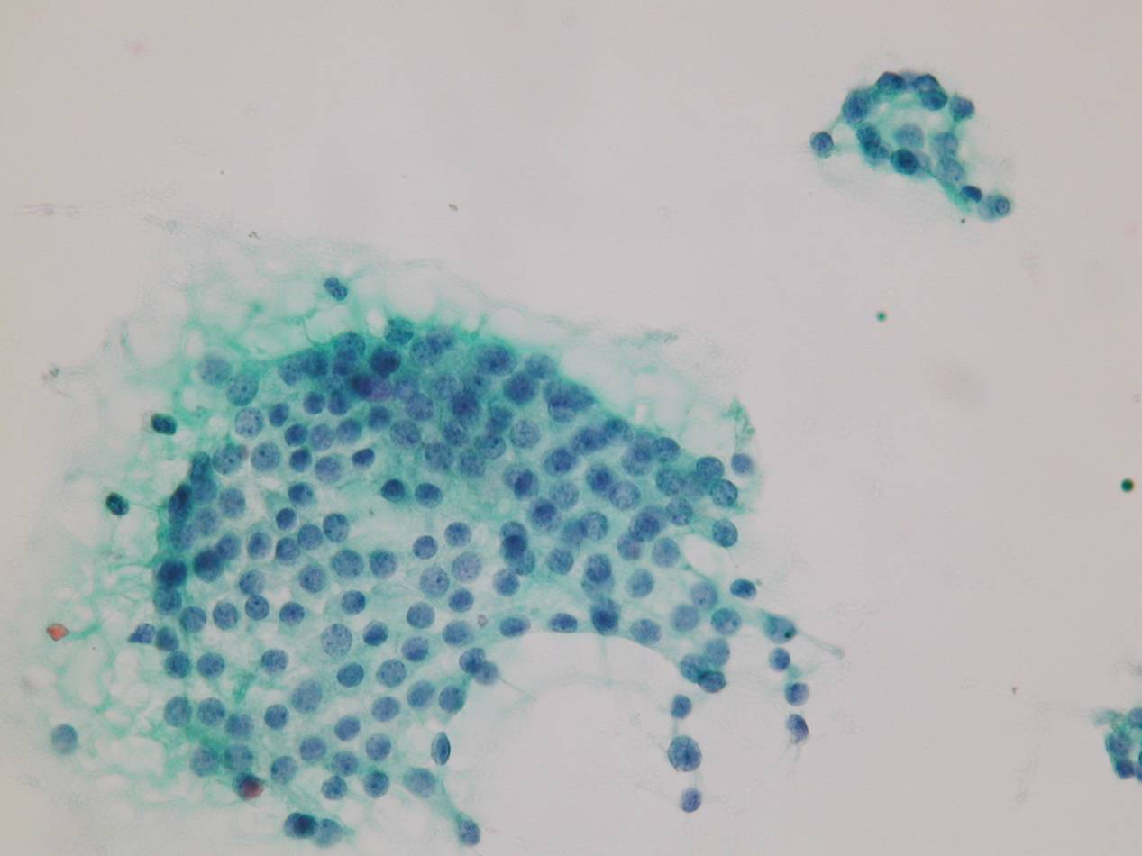
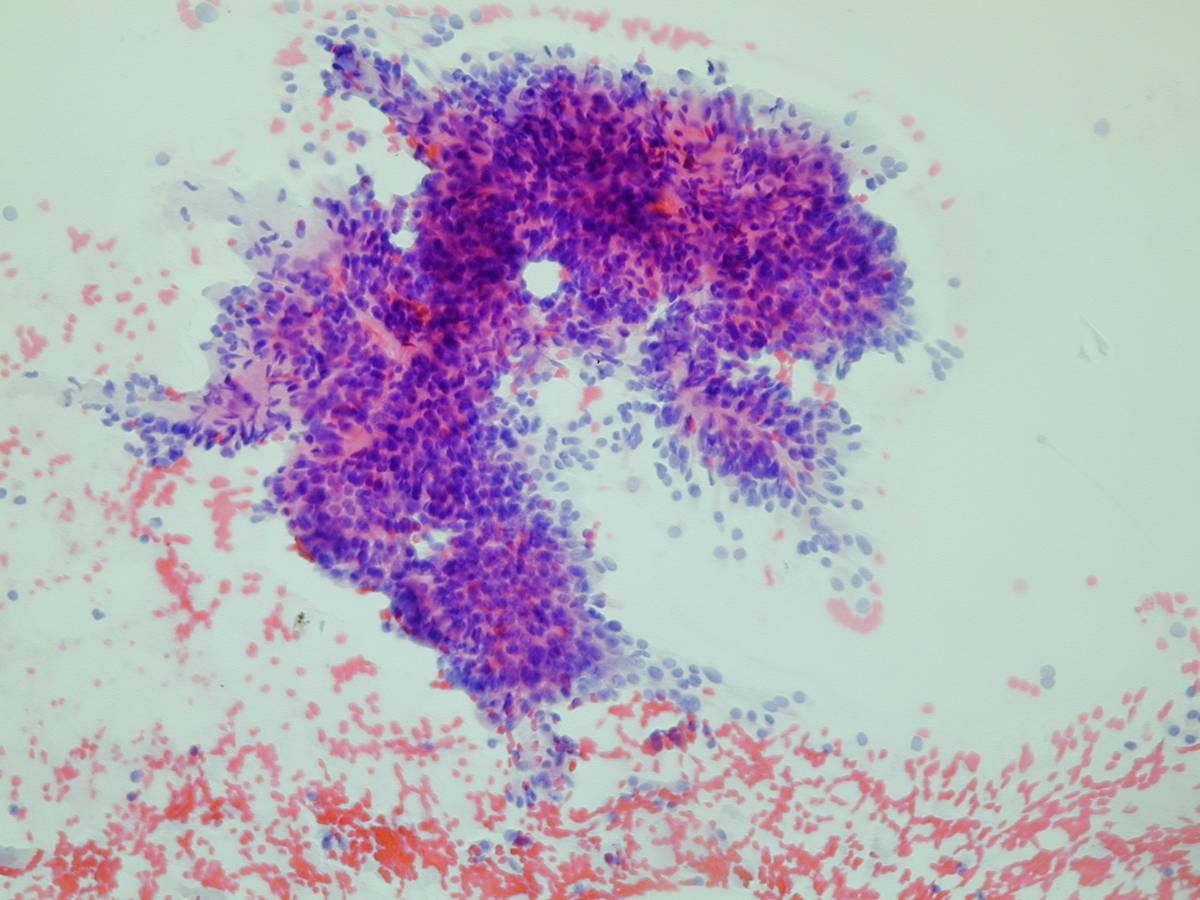
•architecture (monolayers, crowded clusters, sheets, macro/microfollicles, papillary clusters, isolated cells)
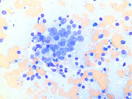
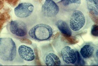
A papillary cluster. •presence of bare nuclei
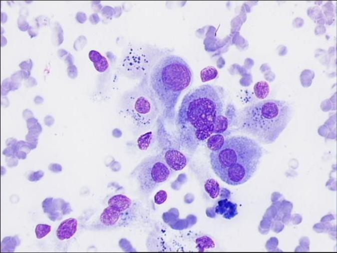
•cytologic features (cytoplasm, nuclei)
In neoplastic lesions the specimens are usually highly cellular. Flat sheets are common in goitre and macrofollicular adenomas, but can also be present in carcinomas. Macrofollicles are usually associated with multinodular goitre and macrofollicular adenomas. A predominance of microfollicles can be suggestive of a follicular neoplasm. Papillary clusters, with cells lining a fibrovascular core, are characteristic of papillary carcinoma. Smears with a high ratio of colloid to follicular cells usually indicate a benign nodule.
Cytologic features:
- cytoplasm (amount, staining)
- chromatin pattern
- nuclear membrane (smooth or irregular)
- nuclear groovings and pseudoinclusions