| Introduction |
| Technical aspects |
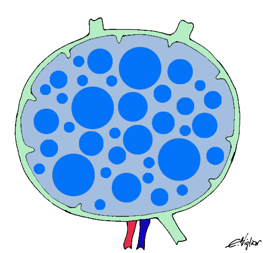
| Normal lymph node histology and cytology |
| Reactive lymph node |
| Hodgkin Lymphoma |
| Non Hodgkin lymphoma |
| Metastases |
NON HODGKIN LYMPHOMA (NHL)
General features
NHL is a heterogeneous group of neoplasms which represent 2-3% of all tumors in the Western World. They encompass both the slowest and the fastest growing tumors, ranging from chronic lymphatic leukemia/small lymphocytic lymphoma which, in the early stages, may be left untreated (watch and wait), to lymphoblastic lymphoma, which require timely and intense therapies. Moreover some entities are well identified by clinical, phenotypical, genetic and molecular profiles, as is generally the case for small-cell lymphomas. Others, such as diffuse, large, B-cell lymphoma, seem to be a container for different molecular phenotypical and clinical entities. Clinical signs, when present, are weight loss, itch, fever, weakness, cough or the mediastinal syndrome if the mediastinum is involved, or only lymph nodal enlargement. The site of the lymph nodes involved is quite variable, although anterior cervical nodes are the ones most frequently affected, followed by supra-clavear and axillary nodes. The NHL classification has been reformulated several times, reflecting the huge amount of knowledge gathered in the field of immunology and genetics of lymphomas.
Back to topNHL Classification
The World Health Organization Classification of Lymphomas (2001)
B-CELL NEOPLASMS
Precursor B cell neoplasm
- Precursor B-lymphoblastic leukemia/lymphoma
- Precursor B-cell acute lymphoblastic leukemia
Mature (peripheral) B-cell neoplasms
- B-cell chronic lymphocytic leukemia/small lymphocytic lymphoma (SLL/CLL)
- B-cell prolymphocytic leukemia Lymphoplasmacytic lymphoma (LPCL)
- Splenic marginal zone B-cell lymphoma (with/without villous lymphocytes)
- Hairy cell leukemia
- Plasma cell myeloma/plasmacytoma
- Extranodal marginal zone B-cell lymphoma of MALT type (MALT)
- Nodal marginal zone B-cell lymphoma (with/without monocytoid Bcells) (MZL)
- Follicular lymphoma (FL)
- Mantle-cell lymphoma (MCL)
- Diffuse large B-cell lymphoma (DLBCL)
- Burkitt lymphoma/Burkitt cell leukemia (BL)
T-CELL AND NK-CELL NEOPLASMS
Precursor T-cell neoplasm
- Precursor T-lymphoblastic lymphoma/leukemia
- Precursor T-cell acute lymphoblastic leukemia
Mature (peripheral) T-cell neoplasms
- T-cell prolymphocytic leukemia
- T-cell granular lymphocytic leukemia
- NK-cell lymphomaleukemia (NKL)
- Adult T-cell lymphoma/leukemia (HTLV-1 positive)
- Extranodal NK/T-cell lymphoma, nasal type
- Enteropathy-type T-cell lymphoma
- Hepatosplenic gamma-delta T-cell lymphoma
- Subcutaneous panniculities-like T-cell lymphoma
- Mycosis fungoides/Sezarysyndrome
- Anaplastic large-cell lymphoma, T/null cell, primary cutaneous type
- Peripheral T-cell lymphoma, not otherwise characterized (PTL)
- Angioimmunoblastic T-cell lymphoma
- Anaplastic large-cell lymphoma, T/null cell, primary systemic type
Back to topRole of cytology
Back to top
- The cytological diagnosis of NHL is hampered by objective difficulties. As for other neoplasms, many NHLs lack significant nuclear atypia because they are the clonal expansion of lymphoid cells in specific stages of maturation. Moreover these cells may be intermingled with different amounts of benign reactive lymphocytes, as in T-rich B-cell NHL, where reactive lymphocytes may prevail over neoplastic ones.
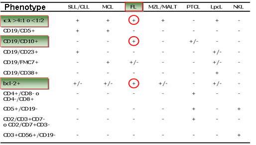
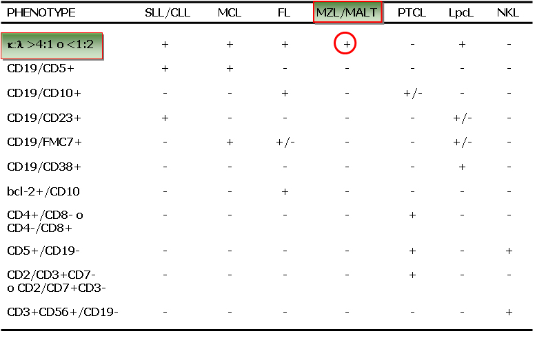
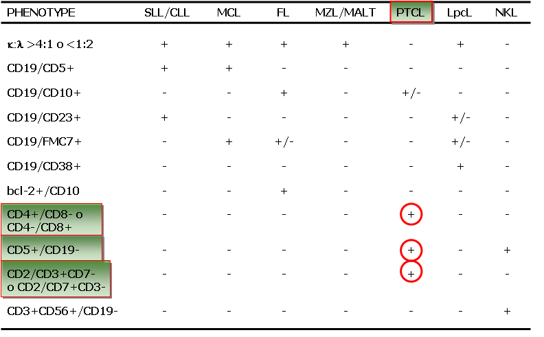
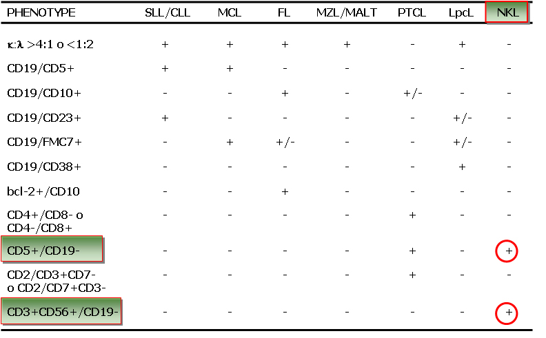
- In practice, the cytological diagnosis of NHL is determined by a combination of microscopic features and ancillary techniques. Differentiated B-cell lymphomas generally show a more monomorphous cell pattern, but a definitive diagnosis may be made only by light chain clonality (κ/λ >4:1 or <1:1) or IGH rearrangement. Attention should be paid to quantitatively small unbalancements, especially if detected by FC (less than 20% of gated cells). In fact, small unbalancements, in the absence of lymphoma, may occur in reactive lymph nodes mainly in cases of autoimmune or immunodeficiency syndromes. Other than light chain restriction, there are other phenotypic patterns, mainly assessed by FC, which define a lymphoid cell population as B-lymphoma, and are frequently sub-type specific: CD5/CD19 co-expression (CLL/SLL or MCL); CD10/Bcl-2 co-expression (FL), CD10+ in all the gated cells, combined with a specific cytological pattern (BL, FL). T-cell lymphoma are more complex; peripheral T-cell generally express CD2/3/7 and alternatively CD4 or CD8. Loss of one antigen (often CD7) may suggest a clonal proliferation. CD3/CD56 over-expression is indicative of NK lymphoma.
- Large cell B-cell lymphoma (DLBCL), generally presents more evident nuclear atypia in all its microscopic and phenotypic variants (centroblastic, immunoblasic, anaplastic). In these cases cytological features and phenotypic assessment can be diagnostic (CD19+, aberrant phenotypes by FC, ALK+,CD30-, EMA+ in DLBCL ALK+ by ICC).
- Cytological features combined with ancillary techniques allow a correct diagnosis and classification in most cases. However cytopathologists should be aware that objective limitations still exist in both the diagnosis and classification of NHL. When a defined diagnosis of B NHL has been assessed, a possible sub-classification may be attempted combining cytological features and phenotypic patterns using ICC or FC.
- In many institution the cytological diagnosis of NHL is accepted without histological control in cases of relapses and in peculiar primary cases; conversely the histological confirmation is necessary for primary diagnoses and in all cases where cytological, clinical and instrumental findings appear contradictory. In addition to repeating the test, surgical excision and histological examination should always be included in the cytological reports of all doubtful cases.
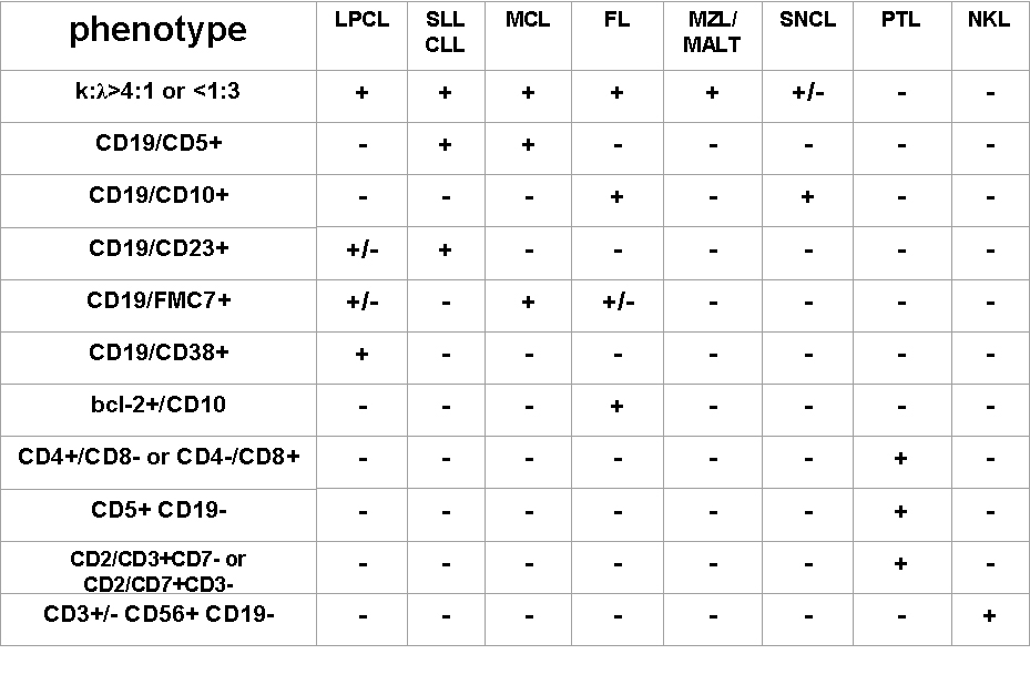
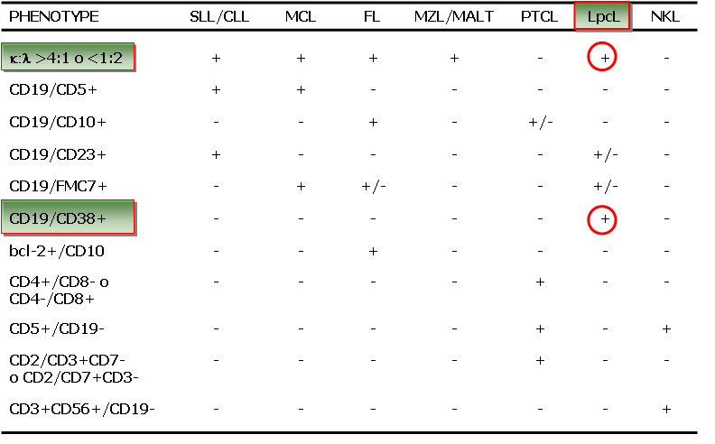
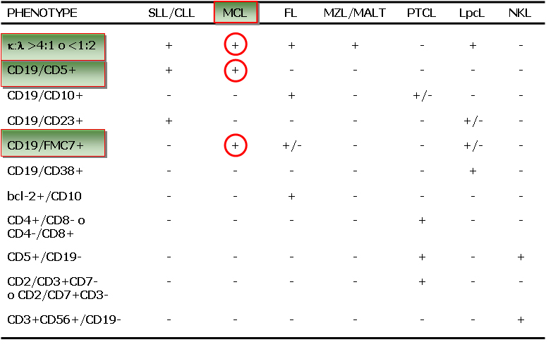
ICC-FC sub-classification of small/medium cell size NHL
Back to top Diagnostic criteria for NHL
Chronic lymphatic leukaemia/small lymphocytic lymphoma (CLL/SLL)
- Incidence: 7-8% of all NHL.
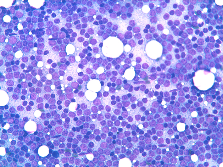
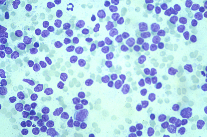
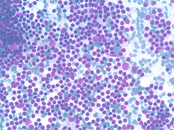
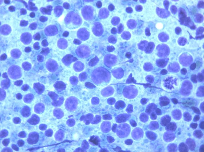
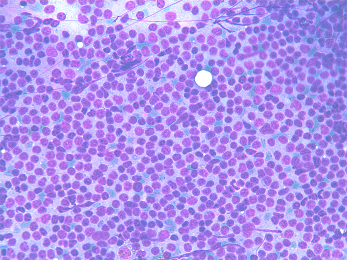
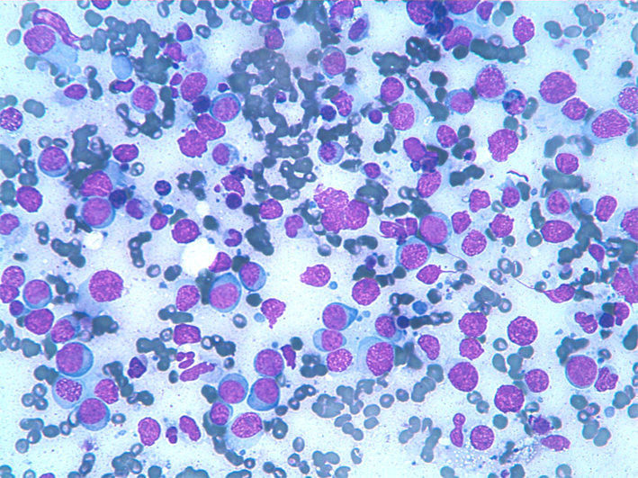
- Cytology: Smears show monotonous, dispersed cell populations composed of small lymphocytes with clumped chromatin and occasional, small, nucleoli; mitoses are absent or rare. Accelerate phases of CLL/SLL show larger, nucleolated, cells (paraimmunoblasts) and mitoses, and should not be confused with Richters syndrome, which is a large-cell histiocytic lymphoma arising on a CLL/SLL background.
- ICC: light chain restriction, CD5+, CD20+, CD23+; Ki 67 is positive in a small percentage of the cells.
- FC: CD5/CD19+, CD19/CD23+, CD19+/CD10-, κ or λ light chain restriction.
- FISH: trisomy 12, 11q22 (ATM), 12, 13q14.3, 13q34.3 (LAMP1), and 17p13.1 (p53) as prognostic markers.
- Differential diagnosis: Non-specific hyperplasia ML, FL, MZL.
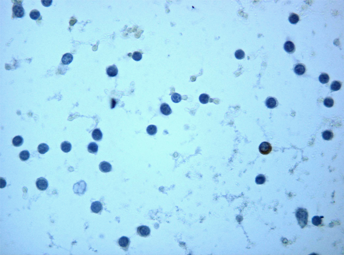
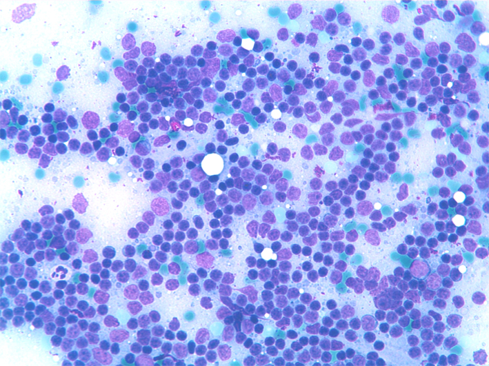
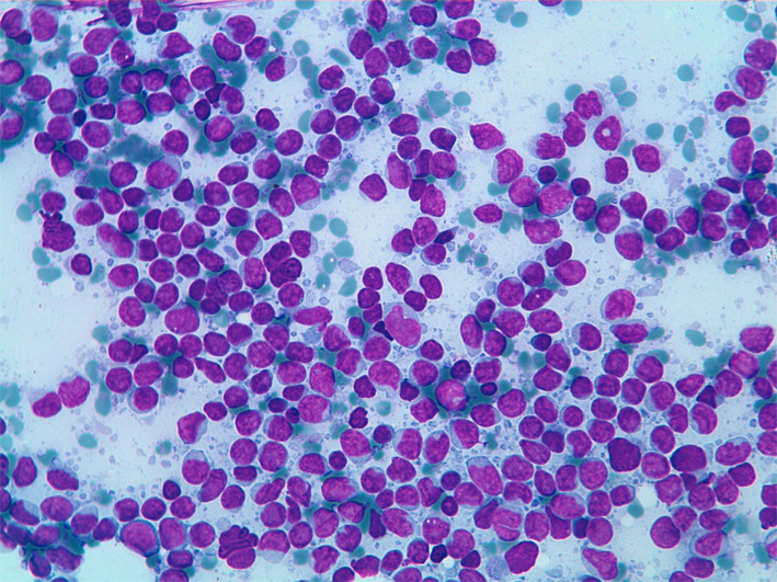
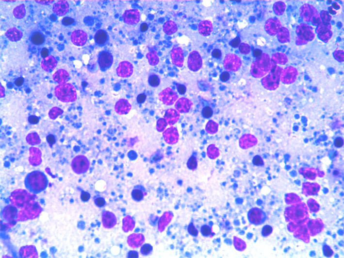
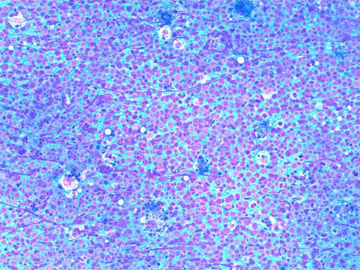
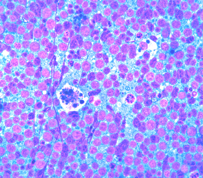
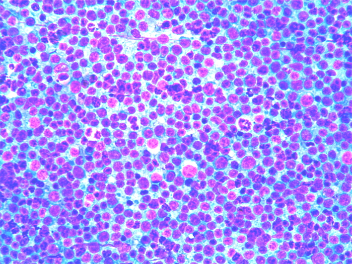
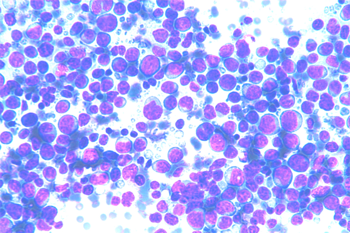
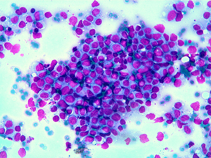
CLL/SLL: A monotonous monomorphous cell population of small lymphocytes with clumped chromatin and occasional, small, nucleoli.
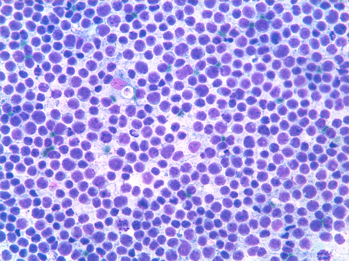
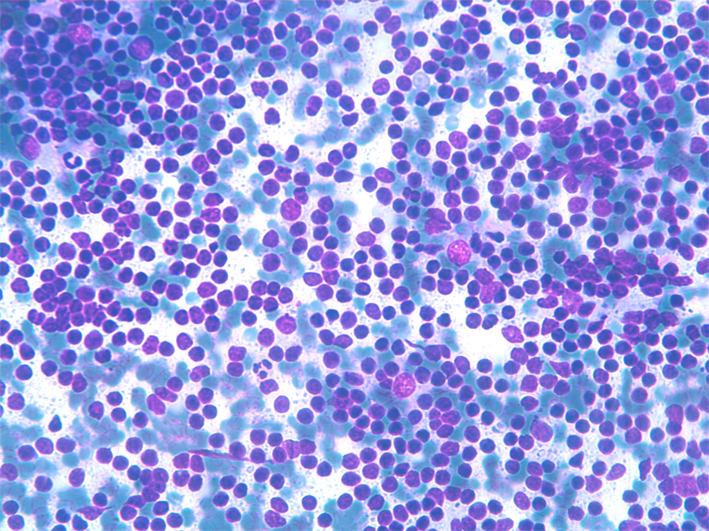
SLL/CLL smears shows a monomorphic, dispersed cell population composed by small lymphocytes and occasional larger nuclei. Small cytoplasm fragments (glandular bodies) are present in the background.
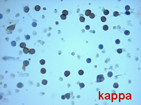
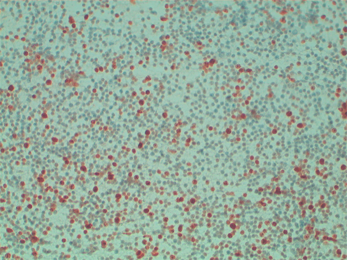
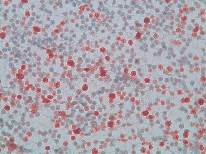
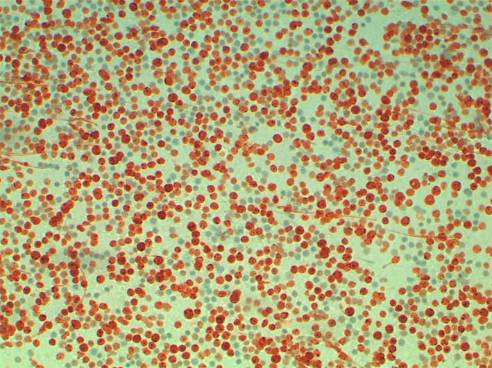
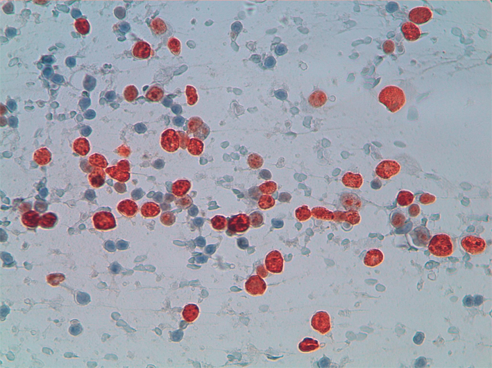
ICC of SLL/CLL on cytospins showing negativity for lambda and positivity for kappa light chains (peroxidase/anti peroxidase immunostain)
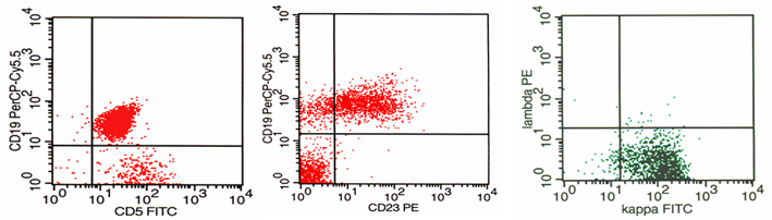
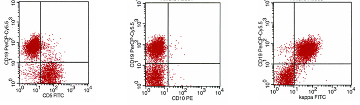
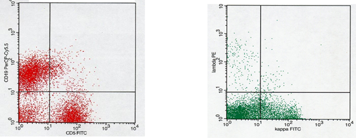
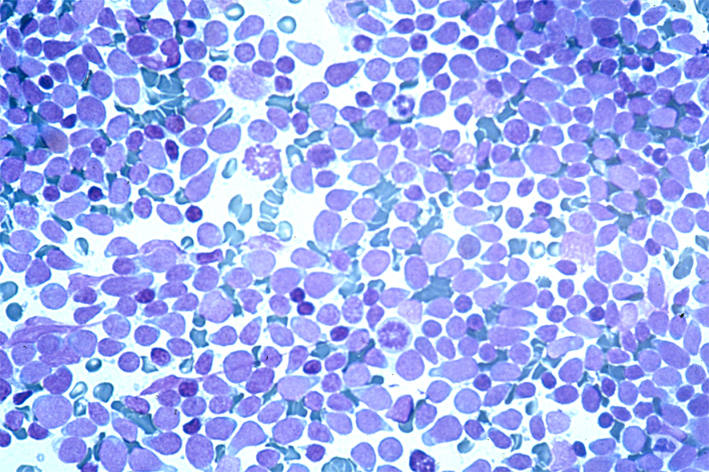
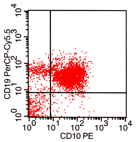
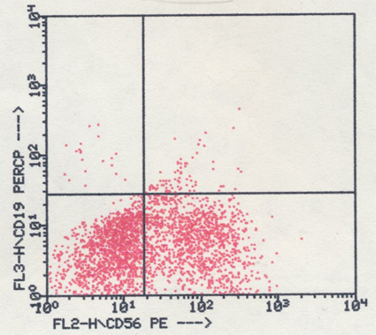
CLL/SLL: FNA smear and corresponding FC showing CD19/CD5 and CD19/CD23 co-expression and kappa light chain restriction.Lymphoplasmacytoid Lymphoma, Immunocytoma (LPCL)
- Incidence: 1-2% of all NHL.
- Cytology: dispersed cell population composed of small lymphocytes with plasmacytoid differentiation; mature plasma cells are also present.
- ICC: CD20+, CD5-,CD10-, CD23-, Ki67+ in a variable percentage (10-20%) of cells.
- FC: CD5-/CD19+, CD19+/CD23+, CD19+/CD10-, CD19+/CD38+, κ or λ light chain restriction.
- Differential diagnosis: Non-specific hyperplasia, SLL/CLL.
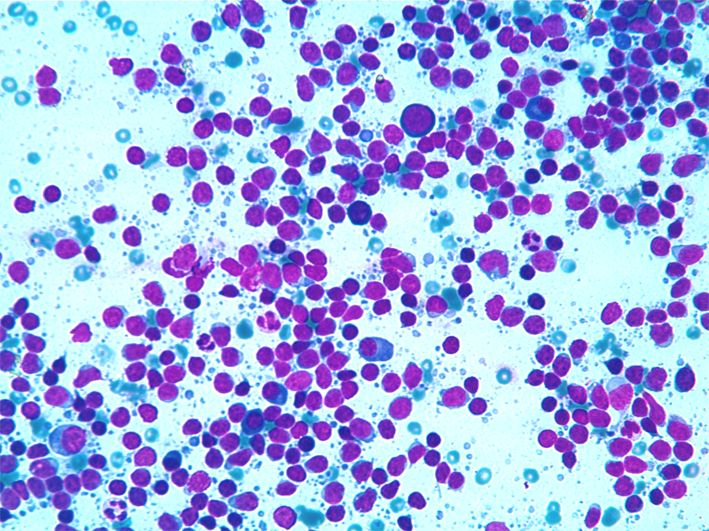
LPCL dispersed cell population composed by small lymphocytes with clumped chromatin and plasmacytoid differentiation
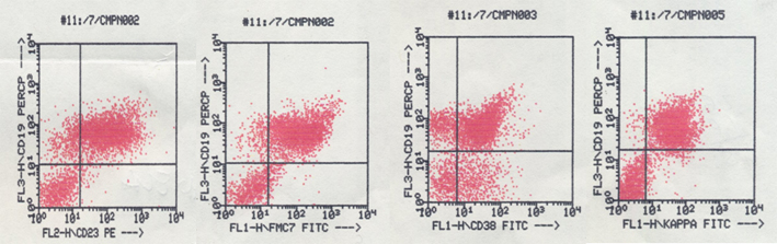
LPCL FC showing co-expression of CD19/CD23, CD19/FMC7, CD19/CD38 and light chain restriction.Mantle cell lymphoma (MCL)
Back to top
- Incidence: 7-10% of all NHL.
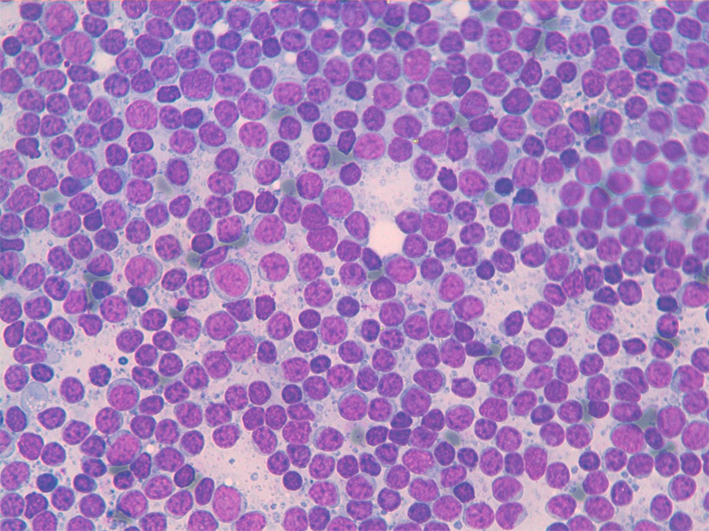
- Cytology: dispersed cell population composed of small/medium-size lymphocytes with irregular nuclei and a thin rim of pale cytoplasm. Nuclei may have slightly irregular shape, clumped chromatin and small nucleoli, or may be cleaved. Mitoses are scanty whereas they may be numerous in the blastoid variant
- ICC. CD20+, CD5+,CD10-, CD23-, Ki67+ in a variable percentage (10-20%) of the cells.
- FC: CD5+/CD19+, CD19/FMC7+, CD19+/CD23-, CD19+/CD10-, κ or λ light chain restriction.
- FISH: t(11;14)(q13;q32).
- Differential diagnosis: Non-specific hyperplasia, SLL/CLL,FL, MZL.
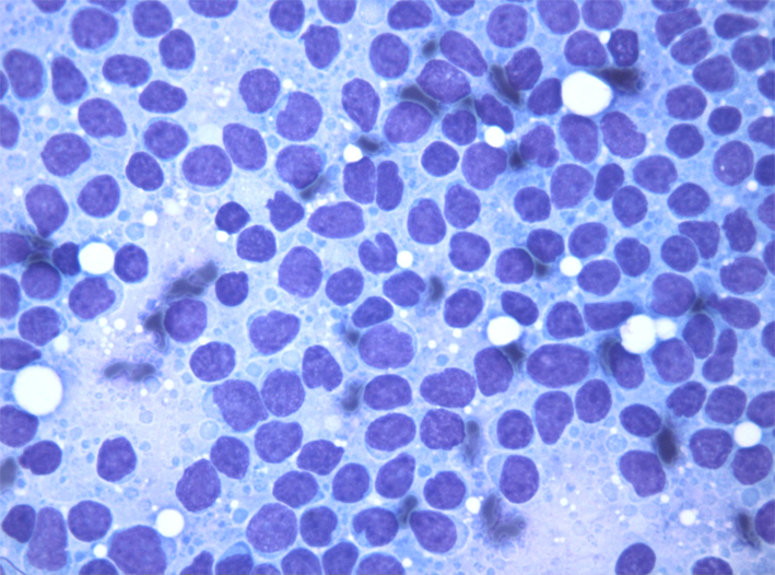
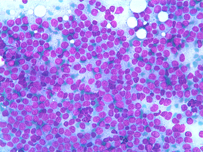
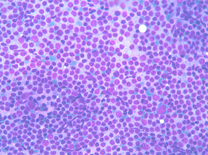
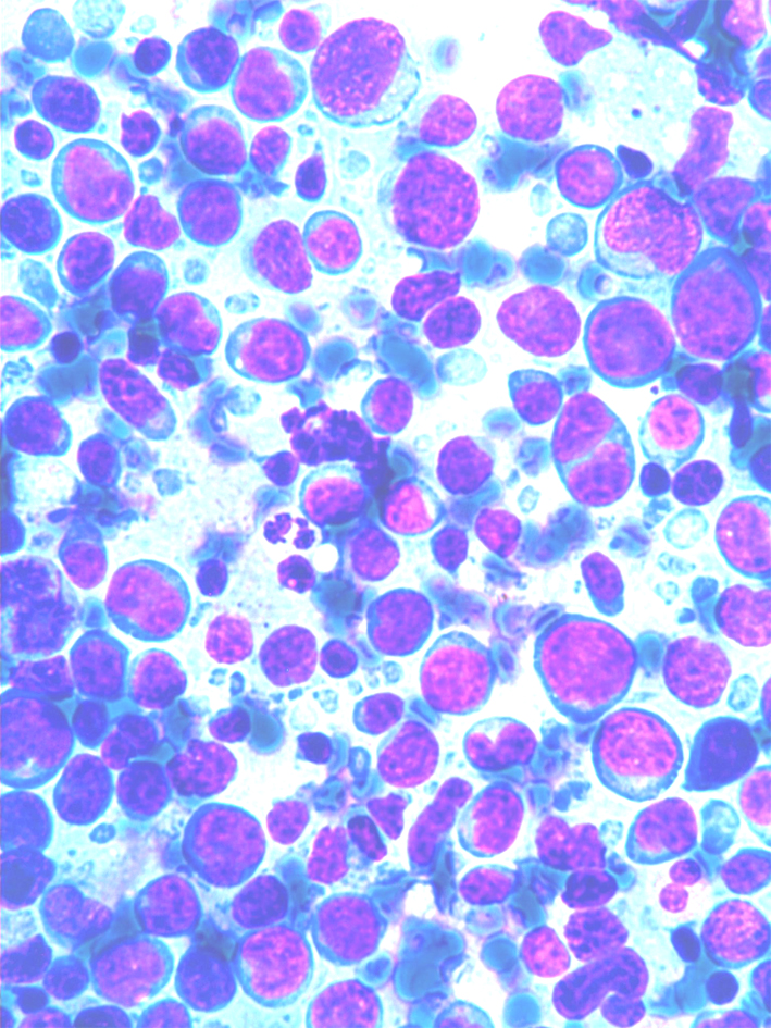
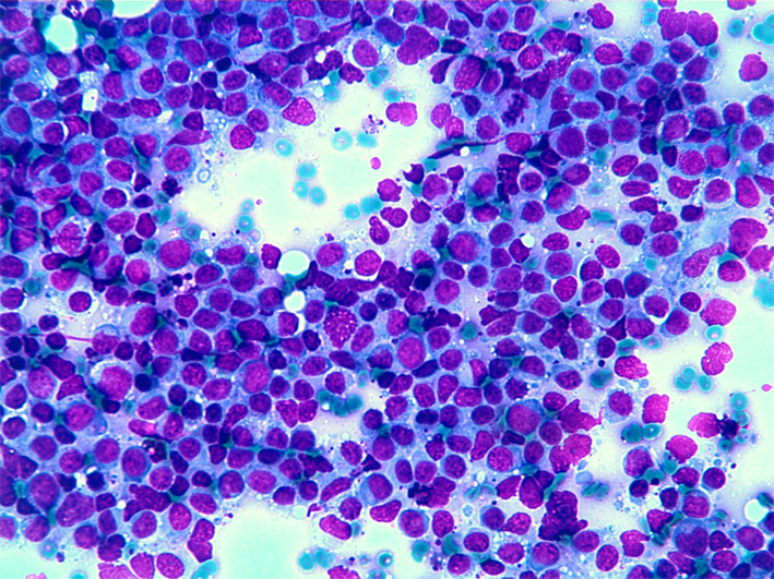
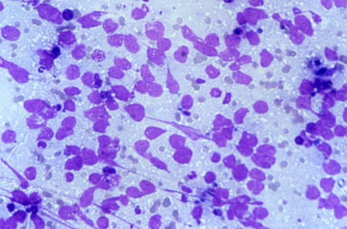
MCL: dispersed cell population composed by medium-size lymphocytes. Nuclei may be slightly irregular in shape or cleaved and have clumped chromatin and small nucleoli.
MCL: dispersed cell population composed by medium-size lymphocytes with irregular nuclei. Nuclei are slightly irregular in shape or cleaved and have clumped chromatin and small nucleoli.
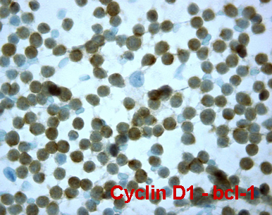
MCL: nuclear positivity for cyclin D1.
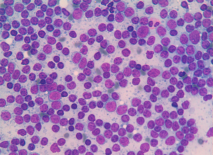
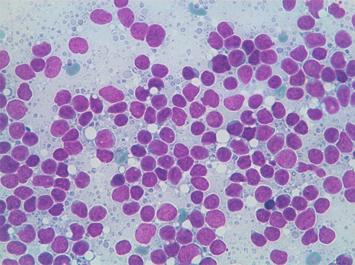
MCL: Medium-size lymphocytes with irregular nuclei and clumped chromatin. Frequent mitoses may be seen.
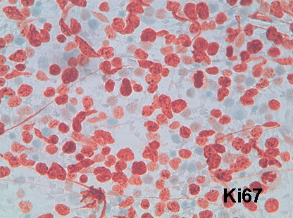
MCL: Ki67 immunostain may shows nuclear positivity in a high percentage of cells.
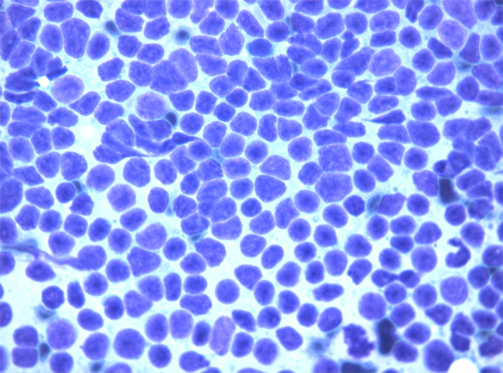
MCL: Medium-size lymphocytes with irregular nuclei and clumped chromatin. Nuclei may show a kind of moulded pattern.
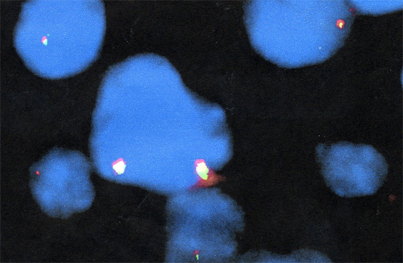
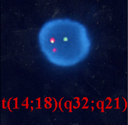
MCL: FISH demonstration of t(11;14)(q13;q32): locus 14q32 (IGH) is labelled in green and locus 11q13 (CNDD1/PRAD1) is labelled in orange. Note fusion signals (yellow) in which the green signals overlap the orange signals.
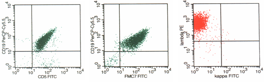
FC of MCL showing CD19/CD5 and CD19/FMC7 co-expression and lambda light chain restriction. Follicular lymphoma (FL)
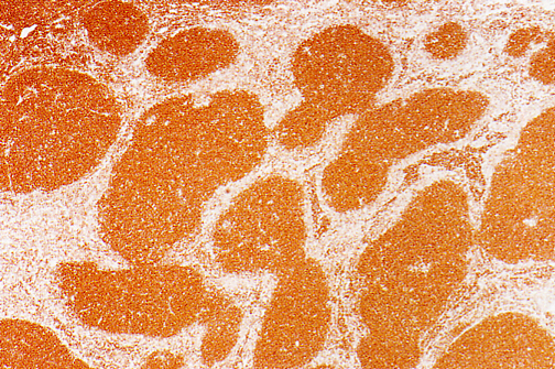
Follicular lymphoma (FL): cytology
- Incidence: 22-30% of all NHL.
- Cytology: dispersed cell population composed of a spectrum of medium- to large-size lymphocytes, with irregular nuclei and a thin rim of pale cytoplasm. Nuclei may have slightly irregular shape, or be cleaved with clumped chromatin and inconspicuous nucleoli in grade I, to large nuclei with dispersed chromatin and two or more marginal nucleoli in grades II and III.
- ICC: CD20+,CD5-,CD10+,CD23-,Ki67+ in a variable percentage of the cells.
- FC: CD5-/CD19+, CD19+/FMC7+, CD19+/CD23-, CD19+/CD10+, CD10/bcl-2+, κ or λ light chain restriction.
- FISH: t(14;18)(q32;q21).
- Differential diagnosis: Non-specific hyperplasia, SLL/CLL,MCL.
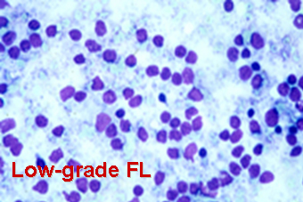
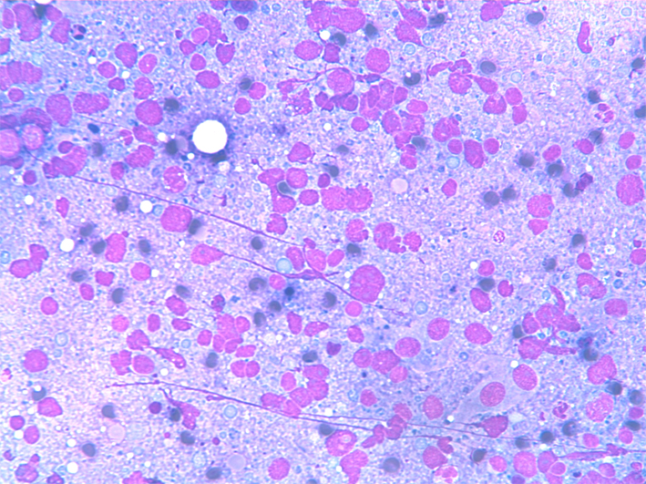
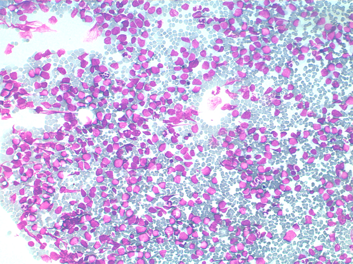
Low-grade FL: a monotonous population of small lymphocytes with clumped chromatin and inconspicuous nucleoli. Scattered large cells are present in the background.
Low-grade FL: Ki-67 show positivity in a relative small percentage of the cells.
Low-grade FL: a monotonous population of small lymphocytes with clumped chromatin and inconspicuous nucleoli. Scattered large cells are present in the background.
Low-grade FL: Ki-67 show positivity in a relative small percentage of the cells.
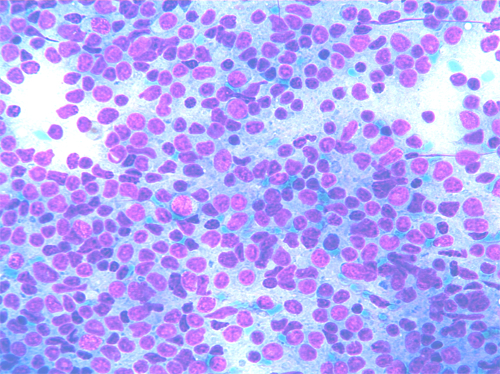
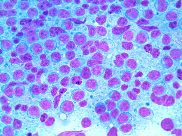
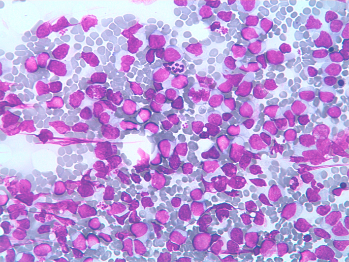
Intermediate-grade FL: a relative monomorphous population of medium-sized lymphocytes with clumped chromatin, nuclear abnormalities and one or more nucleoli.
Intermediate-grade FL: Ki-67 show positivity in a large percentage of the cells.
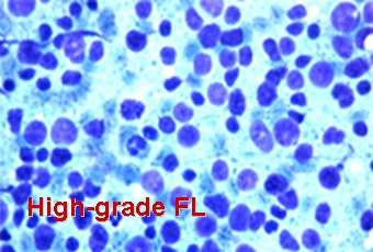
High-grade FL: a relative monomorphous population of large lymphoid cells with dispersed chromatin, nuclear abnormalities and one or more large nucleoli.
High-grade FL: Ki-67 show positivity in almost all the large cells present in the smear.
High-grade FL: a relative polymprphous population of large lymphoid cells with dispersed chromatin, nuclear abnormalities and one or more large nucleoli. Scattered small lymphocytes are present in the background.
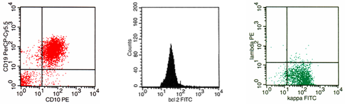
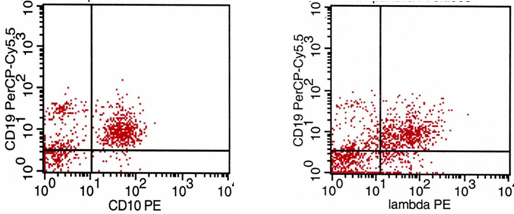
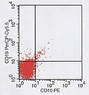
Follicular lymphoma (FL): flow cytometry
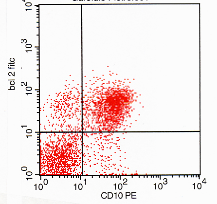
FC of FL showing CD19/CD10 co-expression, bcl-2 overexpression and kappa light chain restriction.
FL: a relative polymorphous population of medium-sized lymphocytes with dispersed chromatin, nuclear abnormalities, and one or more small nucleoli. FC shows CD10/bcl-2 co-expression that reflects the follicles bcl-2 histological positivity.
Follicular lymphoma (FL): FISH
- In cases of FL (CD10/19 co-expression) with dim or no light chains expression, FISH may be helpful in diagnosis by the demonstration of t14-18. Bcl2 locus (18q21,labelled red), overlaps IGH locus (14q32, labelled in green) giving a signal of fusion (yellow).
IGH gene rearrangement in lymphoma with dim or no light chains expression
FC in a lymphoma without light chains expression
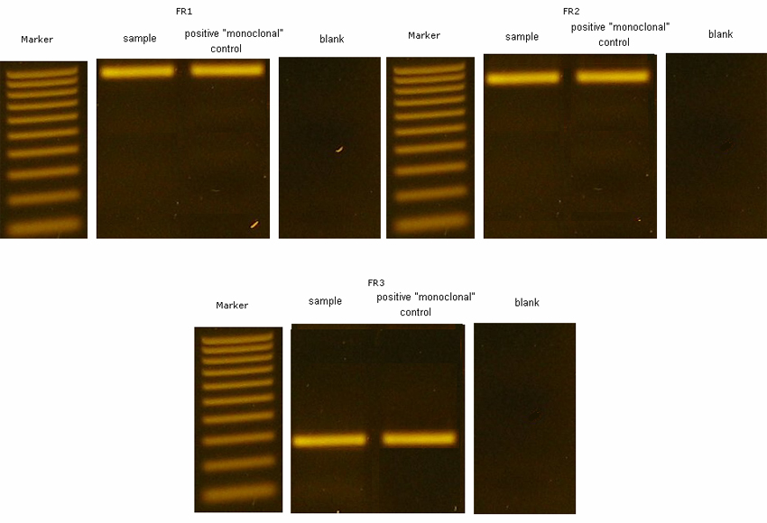
Detection of clonal IGH gene rearrangements: In PCR, rearranged DNA is amplified with a series of consensus primers that are complementary to sequences of variable regions framework 1, framework 2, and framework 3 and to joining regions (JH) of the IGH gene. Monoclonal and polyclonal PCR products can be discerned by their size distributions. There are several methodologies for evaluating PCR amplifications; here conventional electrophoresis in ethidium bromidestained agarose gels (AGE). If a significant population of cells contains the same unique IGH rearrangement, it appears as a well-defined band on each of the AGE here reported. A clonal population of cells is indicated by the presence of a distinct nongermline band, whereas normal or reactive populations have only germline bands and should produce multiple bands (smears). Marginal Zone B-cell Lymphoma (MZL)
- Incidence: 2-3% of all NHL.
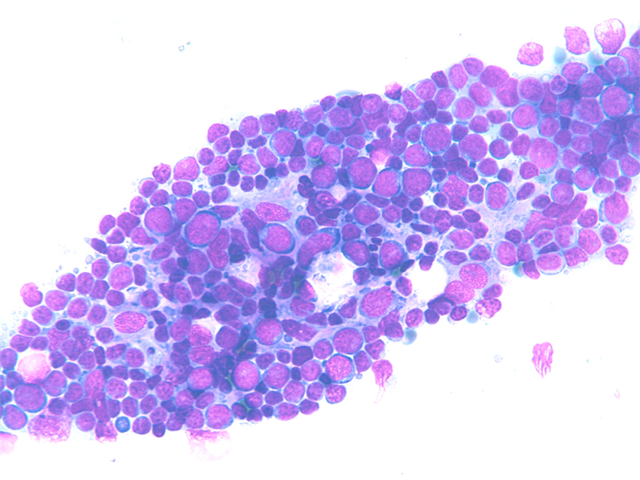
- Cytology: dispersed cell population composed of small lymphocytes with irregular nuclei. Nuclei may have slightly irregular shape, dense chromatin and indistinct nucleoli, or be cleaved. The rim of pale cytoplasm observed in histological sections is rare or undetectable on the smears. Monocytoid cells and plasma cells may be intermingled, mitoses are scanty.
- ICC: CD20+, CD5-,CD10-, CD23-, Ki67+/-.
- FC: CD5-/CD19+, CD19+/FMC7-, CD19+/CD23-, CD19+/CD10-, κ or λ light chain restriction.
- Differential diagnosis: Non-specific hyperplasia, SLL/CLL,MCL, FL.
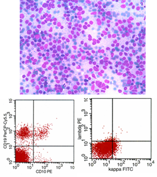
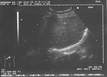
MZL: ultrasound guided FNC of the spleen with diffuse splenomegaly (note the tip of the needle). FNC shows a relative monomorphous population of small lymphocytes with clumped chromatin. FC shows CD5-, CD10-,kappa light chain restriction with CD19/kappa co-expression. Extranodal B-cell lymphoma of mucosa associated lymphoid tissue (MALT)
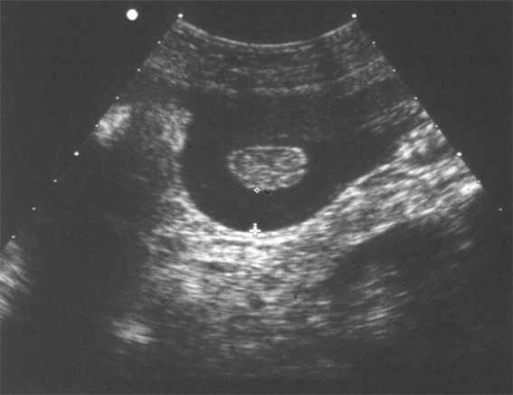
MALT lymphoma: ultrasound guided FNC of an extremely thick bowel wall with former inconclusive endoscopic biopsies. FNC shows atypical lymphoid cells in a necrotic background. FC shows CD5- and kappa light chain restriction. Diffuse large B-cell lymphoma (DLBCL)
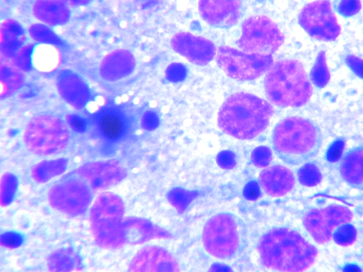
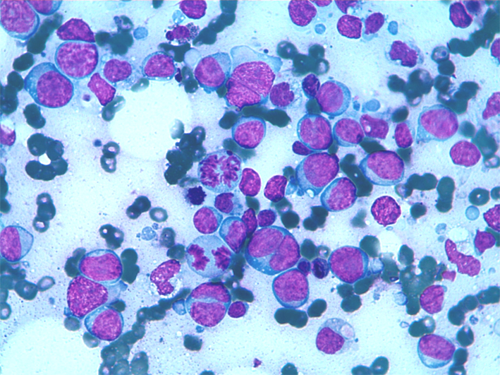
- Incidence: 30-40% of all NHL.
- Cytology: dispersed cell population composed of large lymphoid cells with irregular nuclei, one or more large nucleoli and a wider rim of pale cytoplasm. Morphological variants of DLBCL are centroblastic, immunoblastic T-cell/histiocyte-rich and anaplastic. Nuclei may be slightly irregular in shape or cleaved and have dense chromatin and indistinct nucleoli. Lymphocyte and plasma cells may be intermingled, Mitoses are scanty.
- ICC: CD19+, CD20+, 22+, CD79a+, Ki67+ in a variable percentage (10-20%) of the cells.
- FC: CD5-/CD19+, CD19+/FMC7-, CD19+/CD23-, CD10-, κ or λ light chains restriction.
- DD: HL, metastases.
DLBCL : dispersed cell population composed by large lymphoid cells with irregular nuclei, one or more large nucleoli. Scatterd small lymphocytes are present in the background.
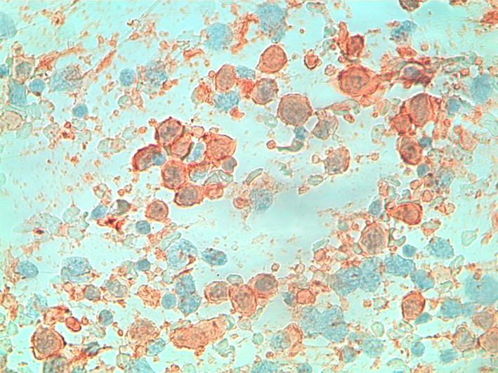
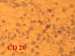
DLBCL : ICC:CD20+.
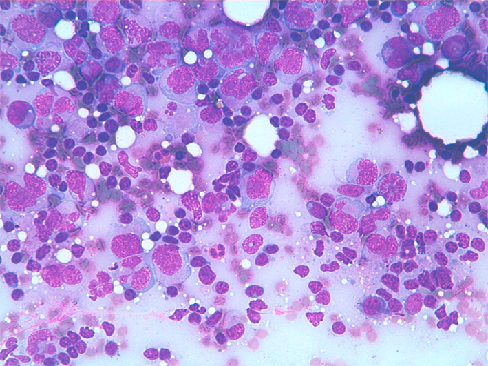
DLBCL : dispersed cell population composed by large lymphoid cells with irregular nuclei, one or more large nucleoli, immature chromatin and nuclear crushes. Cytoplasm may be present or not. Plasma cell myeloma/plasmacytoma
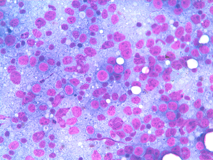
- Incidence: 15% of haematological malignancies.
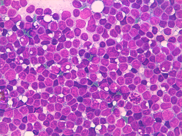
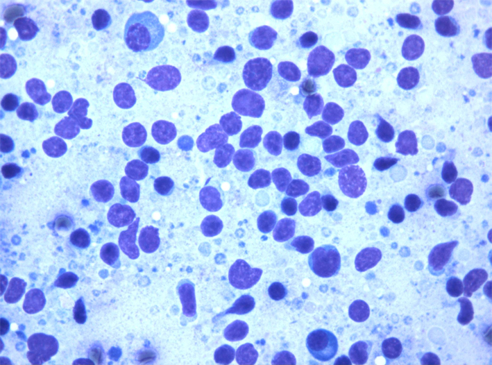
- Cytology: in typical cases show dispersed cell population composed of small/medium-size plasma cells. Cells show one or two nuclei with clumped cartwheel chromatin and small nucleoli. Cytoplasm, when present are well defined with evident perinuclear Golgi apparatus. In poorly differentiated cases nuclei are atypical and may show marked anisonucleosis.
- ICC. CD20-/+, CD5-, CD10-, CD23-, CD38, CD79a, EMA, Ki67+ in a variable percentage.
- FC: CD38+, CD38/56 +/-, CD10-, κ or λ light chain restriction.
- Differential diagnosis: non-haematological tumours in poorly differentiated cases.
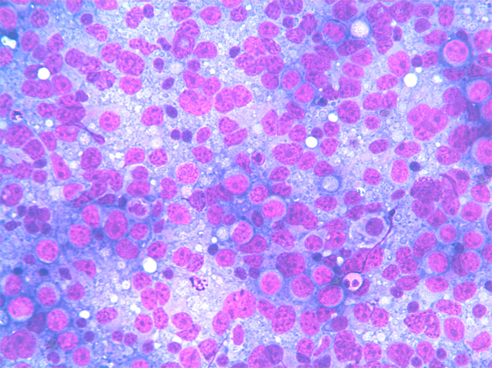
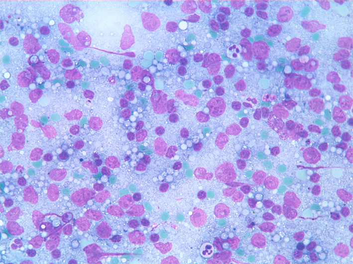
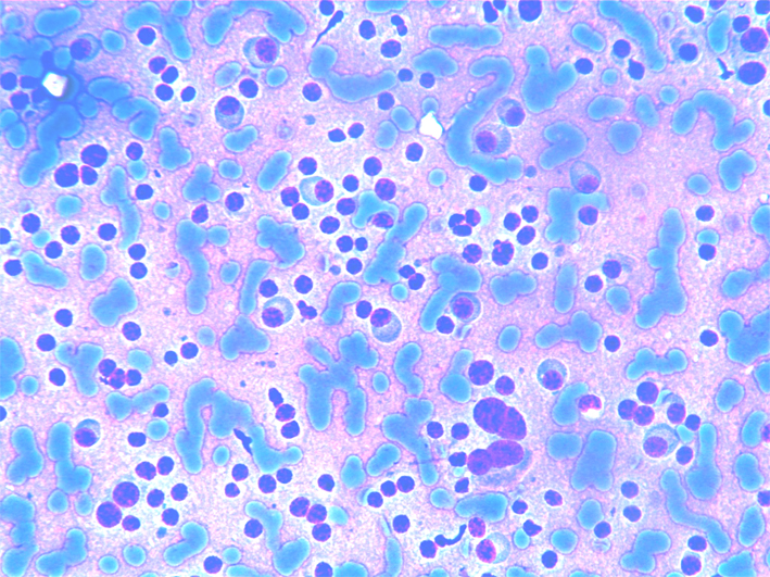
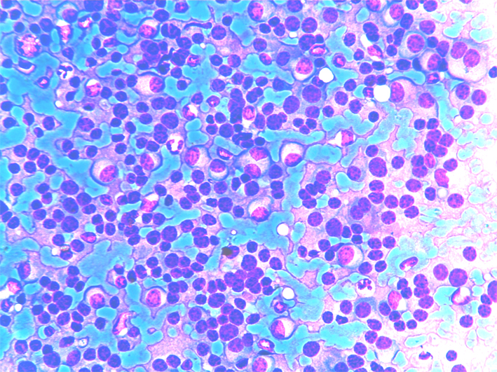
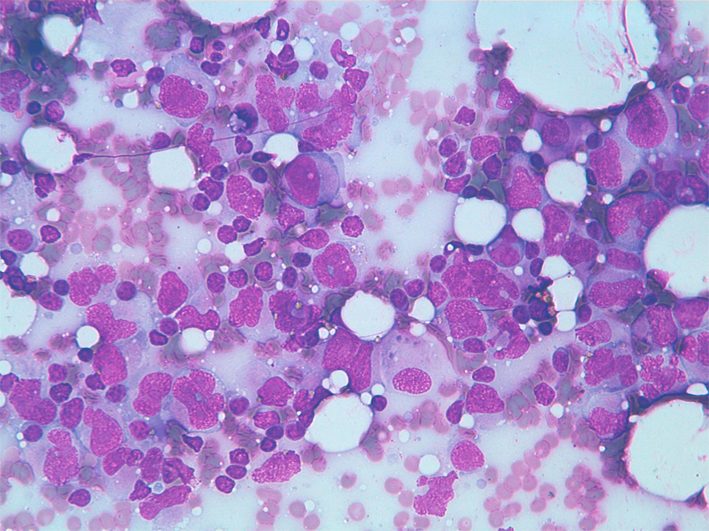
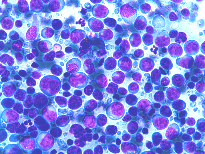
Extramedullary plasmacytoma shows a dispersed cell population of small and medium size plasma cells. Cells show one or two nuclei with clumped cartwheel chromatin and small nucleoli. Cytoplasm, when present are well defined with evident perinuclear Golgi apparatus. Note binucleated cells.
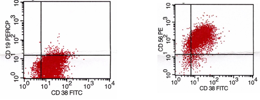
FC of Plasma cell myeloma/plasmacytoma showing CD19-, CD38+, CD38/56 co-expression is observed in 70-80% of the cases multiple myeloma. Lack of CD56 expression has been considered a negative prognostic factor. Burkitt lymphoma (BL)
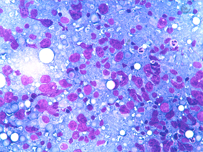
- Incidence: 1-2% of all NHL.
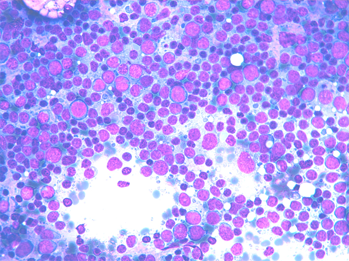
- Cytology: dispersed cell population composed of undifferentiated medium sized lymphocytes with dispersed chromatin and one or more small basophilic nucleoli; the cytoplasm is basophilic and sometimes vacuolated. Immature nuclei are fragile, resulting in frequent nuclear crushes in the smear; a high mitotic index is observed. Scattered macrophages with engulfed cytoplasm may give a starry sky pattern to the smear.
- ICC: CD20+, CD5-, CD10+, Bcl-6+, CD23-, Ki67+ in a high percentage of the cells.
- FC: CD5-/CD19+, CD19+/FMC7-, CD19+/CD23-, CD19+/CD10+, light chain often unexpressed.
- FISH: t(8;14)(8q24;q32).
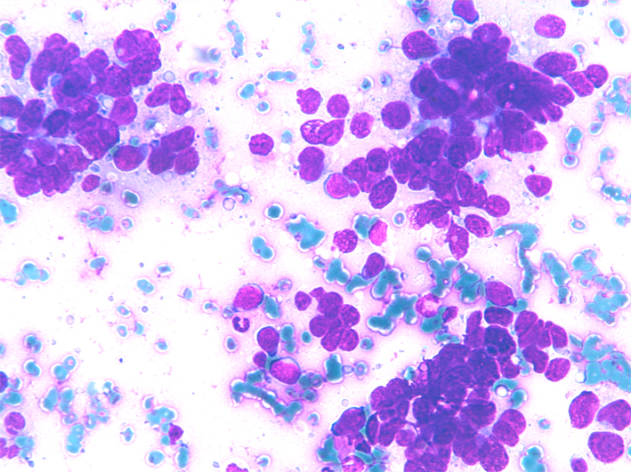
BL: dispersed cell population composed by undifferentiated medium sized lymphocytes with dispersed chromatin and one or more small basophilic nucleoli; Immature nuclei are fragile resulting in frequent nuclear crushes in the smear; an high mitotic index is observed. Scattered macrophages with engulfed cytoplasm may give a starry sky pattern to the smear.
BL: dispersed cell population composed by undifferentiated medium sized lymphocytes with immature chromatin. Numerous mitoses may be observed. FC: almost all the cells shows CD19/CD10 co-expression. Anaplastic large cell lymphoma
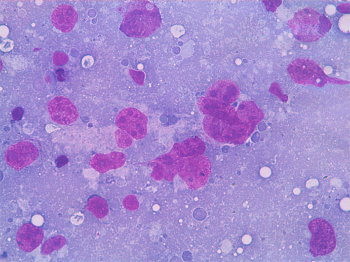
- Incidence: 3% of all NHL.
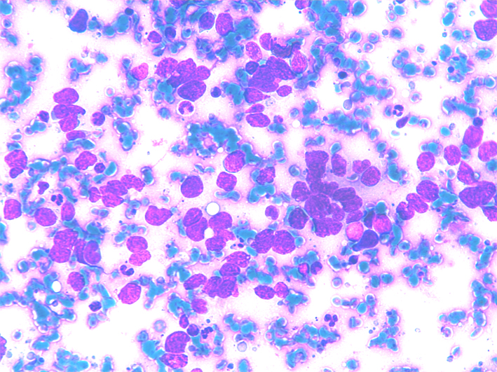
- Cytology: dispersed and polymorphous large cells with very irregular shape. Multilobated, horseshoe bi- and multinuclecleated cells are present. The nuclei have coarse chromatin and larger nucleoli.
- Background is generally polymorphous with mature lymphocytes, granulocytes and eosinophils. Atypical mitoses and necrosis may be seen.
- ICC: CD20-, CD45+ CD15-, CD30++, Ki67+, EMA+, ALK-1+/-.
- FC: CD5+/-, CD19+/-, aberrant phenotypes may be observed.
- FISH: t(2;5)(p23;q35).
- PCR: clonal rearrangement of the T-cell receptor (90%).
- Differential diagnosis: DLBCL, HL, metastases.
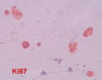
Large anaplastic, nucleolated cells, Ki67+ with small lymphocytes and granulocytes in the background.
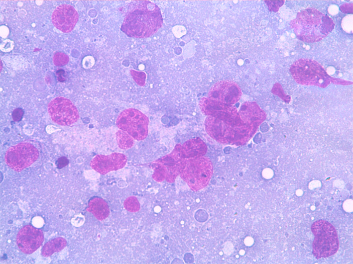
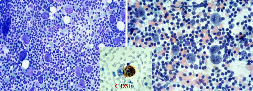
Large anaplastic, nucleolated cells, 30+ interspersed between small lymphocytes Peripheral T-cell lymphoma (PTL)
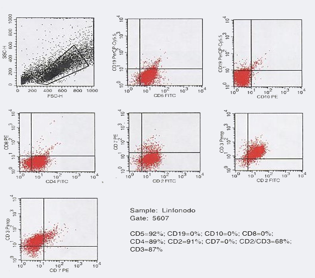
- Incidence: 7-8% of all the NHL.
- Cytology: dispersed and polymorphous medium- or large-sized cells with irregular shape, vesicular nuclei and evident nucleoli. The nuclei have irregular shape, coarse chromatin and one or more large nucleoli. Binucleated cells are frequently observed. The background is generally polymorphous; mature lymphocytes, granulocytes, numerous eosinophils may be present.
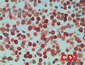
- ICC: CD20-, CD3+, CD4+ CD10-, CD23-, Ki67+ in a variable percentage of the cells.
- FC: CD5+, CD19-, CD10-, CD2+ CD3+ CD7-.
- PCR: TC receptor rearranged in most cases.
- Differential diagnosis: B-cell NHL.
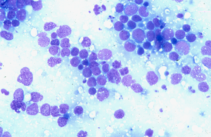
PTL shows a wide spectrum of cytological features: dispersed or loosely aggregated, monomorphic or polymorphous small or medium-sized cells with irregular shape nuclei with evident or inconspicuous nucleoli. The background may be polymorphous with mature lymphocytes granulocytes and/or eosinophils.
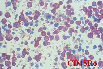
PTL shows polymorphous medium-sized cells with irregular shaped nuclei and small nucleoli. FC: CD19-, CD10-, ICC shows CD20 negativity with few scattered positive cells and diffuse positivity for CD45Ro.
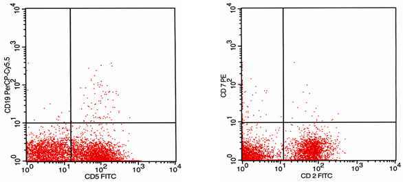
PTL shows polymorphous medium-sized cells with irregular shaped nuclei and scattered small lymphocytes in the background. FC: CD19-, CD5+, CD10-, CD4+, CD8-, CD2+, CD2/3+ CD7-. Loss of CD7 expression is frequently observed in PTL.
PTL shows medium-sized cells poorly differentiated lymphoid cells with irregular shaped nuclei and small nucleoli. ICC shows diffuse positivity for CD45Ro. FC: CD19-, CD5+, CD2+,CD7-.
Precursor T-lymphoblastic lymphoma/leukemia (TLL)
- Incidence: <1% of all the NHL.
- Cytology: dispersed and polymprphous medium-sized undifferentiated cells. Nuclei are round or convoluted with dispersed chromathin and small nucleoli. High mitotic index and nuclear fragility.
- ICC: CD20-, CD3+, CD10-, TDT+, Ki67+ in a high percentage of the cells.
- FC: CD5 +/-, CD19-, CD10-, CD2+ CD3+ CD7-.
- Differential diagnosis: BL.
Dispersed and polymprphous medium-sized poorly differentiated cells. Nuclei are irregular with with clumped chromathin and small nucleoli. Numerpous mitosesmay be seen.
TLL: diffuse CD3+ NK-cell lymphoma/leukemia
- Incidence: <1% of all the NHL.
- Cytology: dispersed and monomorphous small- or medium-sized undifferentiated cells. The nuclei are round or convoluted with dispersed chromatin and small nucleoli. There is also a high mitotic index and nuclear fragility.
- ICC: CD20-, CD3+, CD10-, TDT+, CD56+ Ki67+ in a high percentage of the cells.
- FC: CD56+, CD5 -, CD19-, CD10-, CD2-CD3- CD7-.
- Differential diagnosis: PTL, LLA, TLL.
NK lymphoma: dispersed and monomorphous small and poorly differentiated cells. The nuclei are oval or convoluted with dense chromatin and small nucleoli. Blastic NK-cell lymphoma/leukemia
- Blastic NK lymphoma: dispersed and monomorphous medium-size and poorly differentiated or undifferentiated cells. The nuclei are irregular and fragile with dense chromatin and small nucleoli. Nuclear crushes and atypical mitoses are present. FC is mute except for CD56+