Cervical Cytology
Cervical intraepithelial neoplasia (CIN) invasive squamous carcinoma of the cervix, and atypical squamous cells of undetermined significance (ASCUS)

Morphological characteristics of neoplastic cells

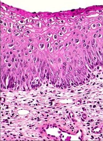
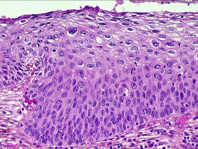
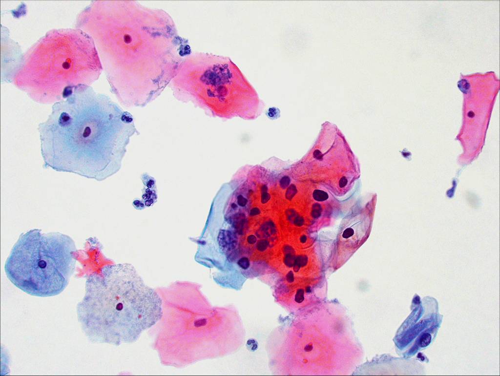
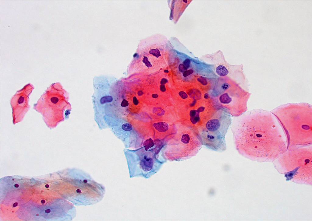
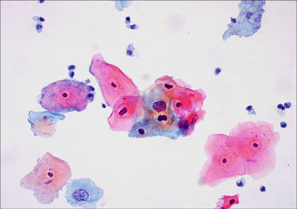
Cervical Intraepithelial Neoplasia(CIN)
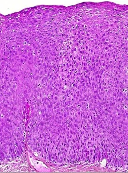
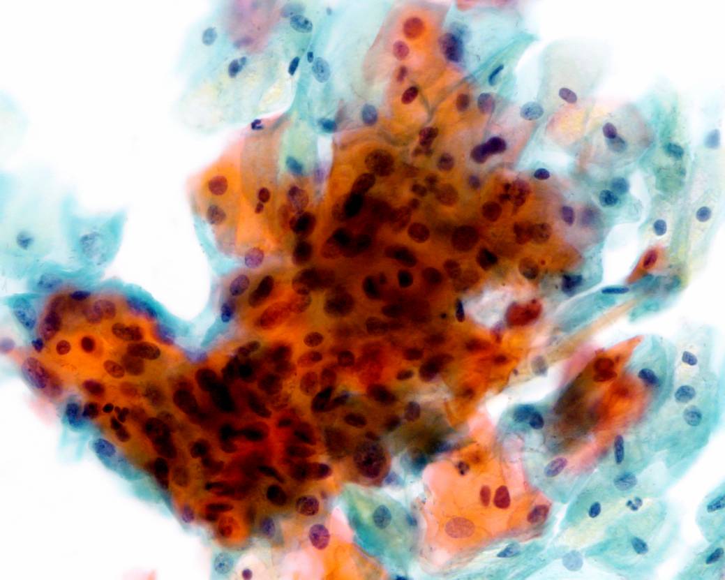
Invasive Squamous Carcinoma
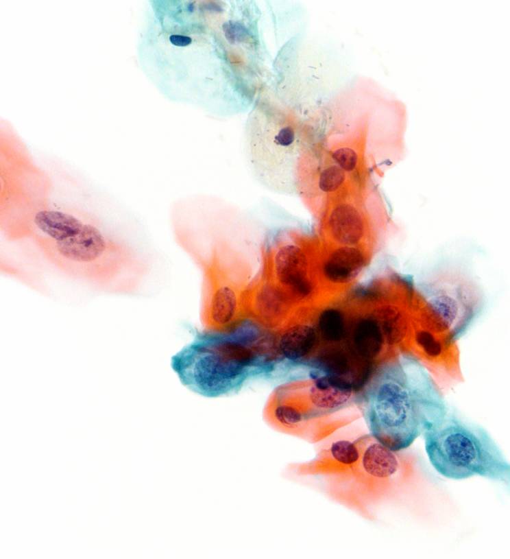
Atypical squamous epithelial cells of uncertain significance (ASCUS)


Morphological & biological characteristics of  CIN

  • In all cases of CIN, the normal epithelium of the cervix is replaced by abnormal epithelial cells showing varying degrees of differentiation.
  • Undifferentiated  neoplastic cells of basaloid or parabasaloid appearance may occupy the whole  thickness of the epithelium (CIN3) or they may differentiate as they approach they approach the surface (CIN1,CIN2)
  • The nuclei of the  undifferentiated cells are large, irregular and vary in size and shape. The chromatin content is increased and abnormal in structure and the nuclear/cytoplasmic ratio is high.
  • As the basaloid  or parabasaloid neoplastic cells differentiate    the amount of cytoplasm increases. Nuclear irregularity, pleomorphism and anisonucleosis persist although there is usually a decrease in nuclear size.
  • Mitotic  activity is no longer confined to the basal layers of the epithelium and atypical mitoses may be seen at any level
  • It is not uncommon for  foci of CIN1,CIN2 and CIN3 to be present in the same cervix
CIN1
CIN2
CIN3

The abnormal cells  found in the smear are derived from the surface of the CIN lesion. The abnormal cells appear  singly or in streaks or in sheets. They can be distinguished from normal cells by the following diagnosis:

  1. Disproportionate nuclear enlargement resulting in alteration of normal nuclear/cytoplasmic ratio.
  2. variation in nuclear size and shape.
  3. irregularity of nuclear outline
  4. hyperchromasia of nucleus  and abnormal chromatin pattern
  5. large irregular and sometimes multiple nuclei (not in CIN1)

The grade of CIN (CIN 1, 2 or 3) can be deduced from the degree of nuclear enlargement and alteration of n/c ratio.


Cytological diagnosis of CIN1 (encompassing LSIL and mild dysplasia)

The Bethesda System definition of CIN 1 (LSIL, mild dysplasia)

  • Nuclear enlargement is three or more times that of a normal intermediate cell nucleus*
  • Nuclear/cytoplasmic ratio may or may not be altered as cytoplasm is usually  abundant and may be keratinised. Cell borders are usually distinct
  • Moderate variation in nuclear size and shape
  • Binucleation or multinucleation are not uncommon
  • Hyperchromasia with uniformly distributed chromatin. The chromatin may appear degenerated or smudged if associated with HPV changes
  • Nucleoli are rare or  inconspicuous
  • Nuclear membrane  may or may not be distinct
  • Cells with a well-defined, optically empty perinuclear halo and peripheral dense cytoplasm (the so called koilocytotic atypia) must also show the above described nuclear abnormalities to be classified as CIN 1(LSIL).

*The WHO cytological definition of CIN 1 requires that the nucleus occupies less than one half the area of the cell.

Cytological presentation of CIN 1 (LSIL).
Cytological diagnosis of CIN2 (encompassing HSIL and moderate dysplasia)
Cytological diagnosis of CIN3 (encompassing HSIL, severe dysplasia and carcinoma in situ)


Back
Next