Cervical Cytology
Cervical intraepithelial neoplasia (CIN) invasive squamous carcinoma of the cervix, and atypical squamous cells of undetermined significance (ASCUS)
| Cervical Intraepithelial Neoplasia(CIN) |
| Invasive Squamous Carcinoma |
| Atypical squamous epithelial cells of uncertain signficance (ASCUS) |
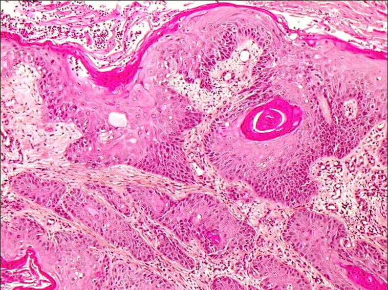
Morphological and biological characteristics of invasive squamous carcinoma
- The pattern of growth of invasive squamous carcinoma of the cervix is very variable. The tumour may be papillary or polypoid or may be flat or ulcerated.
- The tumour may be well differentiated (ie tumour cells may exhibit features of squamous differentiation such as keratin formation) or poorly differentiated (non keratinising). Both keratinised and non keratinised areas are often present in the same tumour although one or other area may be predominant.
- In all forms on squamous carcinoma, the tumour sends out cords of cells into the stroma and surrounding tissue .These appear in histological section as islands of malignant cell surrounded by stroma which is often inflitrated with inflammatory cells.
- Tne tumour cells of invasive squamous carcinoma show much more pleomophism than those of CIN. There is marked variation in nuclear size and shape as well as in the amount and appearance of the cytoplasm. Necrotic cells and cell debris is common as the tumour outgrows its blood supply and infection supervenes.
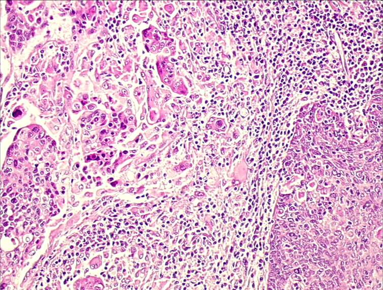
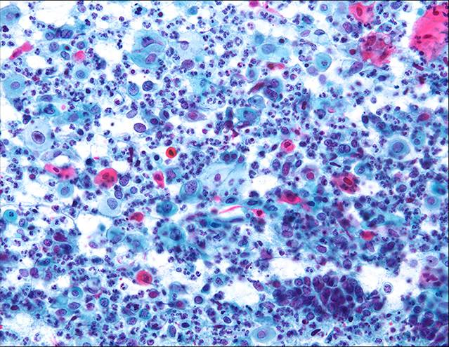
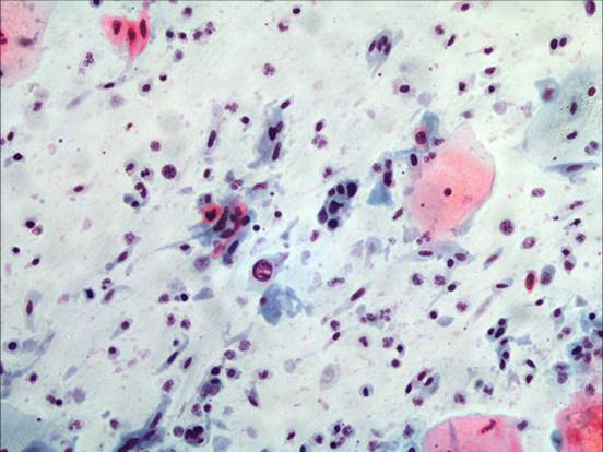
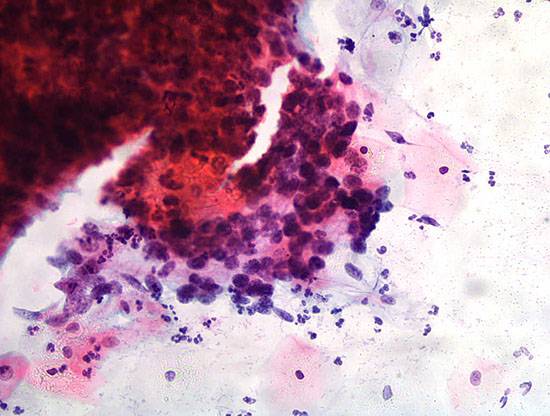
Histological section of keratinising squamous carcinoma Histological section of non keratinising squamous carcinoma Cytological diagnosis of invasive squamous carcinoma
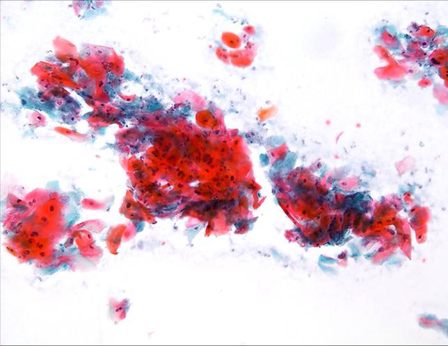
- In keratinising squamous cell carcinomas the tumour cells vary in size and shape. Spindle cells and tadpole cells with highly keratinised orangeophilic cytoplasm are often seen. The nuclei in these cells often show advanced degenerative changes of coagulative necrosis , pyknosis and karryorhexis.
- Anucleate highly keratinised squames with bizzare shapes are not uncommon Keratin plaques or sheets of dyskeratotic or parakeratotic cells may be seen
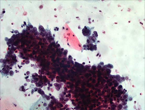
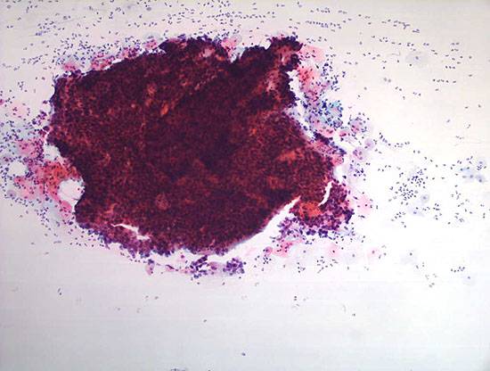
- In non keratinising tumours the cells may be present as single cells or as clusters or syncitial sheets of cells. The nuclear cytoplasmic ratio is abnormal but nucleocytoplasmic ratio is variable. There is a marked nuclear pleomorphism. Nuclei vary in size, shape, chromatin content and chromatin structure. Enlarged irregular, multiple nucleoli may be present. Mitotic figures are sometimes seen
- Intensely staining hyperchromatic fragments composed of poorly differentiated malignant cells may be seen in invasive squamous carcinoma. The fragments appear three dimensional and are composed of epithelial cells with scant cytoplasm and crowded, overlapping large hyperchromatic nuclei which vary in size, shape and chromatin content and structure. Mitotic figures and enlarged multiple nucleoli are often present.
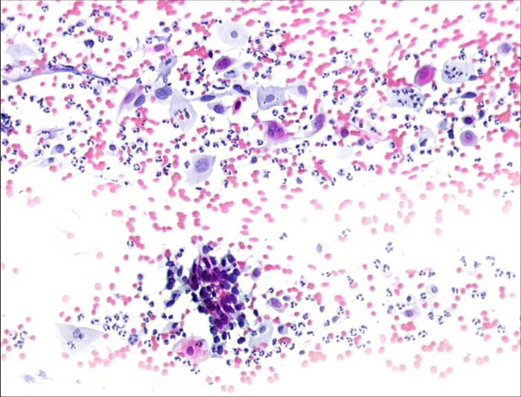
- In advanced lesions the tumour may be ulcerated and infected and the tumour cells embedded in a background of blood, polymorphs and necrotic cells debris (the so called tumour diathesis). Phagocytosis is sometimes seen