Cervical Cytology
Anatomy, physiology and histology of the cervix
Anatomy
Endocervical canal
Ectocervix
Squamocolumnar junction, metaplastic process and Transformation zone


The squamous epithelium of the ectocervix

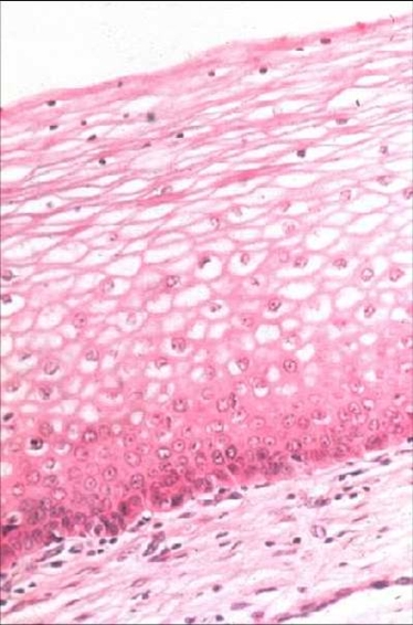
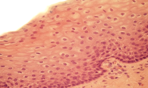
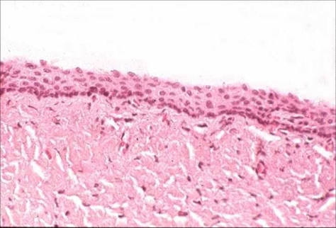
The original squamous epithelium lining the ectocervix is 0.5 mm thick and provides a protective covering for the portio vaginalis of the cervix. It is continuous with the squamous epithelium of the vagina but is less sensitive to hormonal influences than that of the  vagina. Its thickness varies with age and with the menstrual cycle – becoming fully mature under the influence of oestrogen but only semi- mature under the influence of progesterone. In the absence of these hormones e.g. after the menopause, the thickness of the epithelium is greatly reduced.


  • The mature squamous epithelium consisted of 10-20 layers of epithelial cells which can conveniently be divided into three zones.The basal layer (germinal layer)
    1. The superficial layer which is made up of the most mature cells.
    2. The mid or intermediate
    3. The basal layer made up of squamous epithelium
  •  

  • The epithelium is separated from the underlying stroma and  fibromuscular wall of the cervix by a delicate network of fibres which constitutes the basal lamina. The epithelium rests on the underlying connective tissue of the cervix- the stroma. The stroma is comprised mainly of collagen fibres and is densest on the ectocervix and loosely packed around the endocervical glands Smooth muscle and elastic fibres are scarce except high in the endocervical canal in the region of the isthmus. The deep surface of the epithelium is indented in places by stromal papillae which project towards the surface of the epithelium for about one third of its thickness and carries capillary blood vessels into the epithelium.
  • The basal zone : This is a single layer of cylindrical cells 12 μm diameter whose main function is epithelial regeneration. Their oval nuclei display evidence of active cell division such as nucleoli , numerous chromocentres and very occasional mitotic figures. Under normal circumstances the entire process of epithelial regeneration is confined to the basal layers,  the  remaining zones merely reflecting different stages of epithelial cell maturation. Under certain conditions e.g. injury the basal zone may be multi layered giving rise to two or more basal layers. This is termed basal hyperplasia ; above the basal layers  cell maturation is normal.
  • The mid or parabasal zone : This is composed of maturing squamous cells which have more abundant cytoplasm than those of the of the basal zone and hence appear slightly large than the cells of the basal zone. The cells in the upper layers of this zone have more cytoplasm than the deeper zone although the nuclear size remains fairly constant . The epithelial cells of the parabasal zone contain abundant tonofilaments and glycogen granules and the integrity of the epithelium is maintained by desmosomal attachments or intercellular bridges. In consequence this layer is sometimes referred to as the Stratum Spinosum . A granular layer of polyhedral cells containing keratohyaline material is sometimes seen in the uppermost layer of this zone.
  • The superficial layers: These are composed of several layers of loosely attached cells that are broader and thinner than those of the mid zone producing a basket weave pattern. They have small pyknotic nuclei 2-3 μm diameter . Transmission electron microscopy reveals the presence of occasional keratin precursors in the superficial  layers . Full development of keratin does not occur in normal cervical epithelium . However , under certain conditions e.g. vaginal prolapse the superficial cells may synthesize excessive amounts of keratin. Scanning electron microscopy of the superficial layers reveals a  network of  squamous cells which form a mosaic pattern.  The cell surfaces are covered by a network of microridges believed to improve adhesiveness thereby protecting the deeper layers from trauma.


Histology of the squamous epithelium of the cervix  before puberty, during reproductive life and after the menopause:

  • Epithelium at birth and prior to the menarche: At birth,  the cervical epithelium shows some maturation reflecting the actions of maternal hormones in utero. Within a few days the effect wears off and the cervical epithelium becomes thin and immature and is comprised of only a few layers of parabasal type cells. It remains in this state until the menarche.
  • During the Proliferative phase of the menstrual cycle: The epithelium  responds to the influence of oestrogen by  complete maturation. It is composed of 18 - 20 layers of large squamous cells containing keratin precursors which protects the cervix from the vaginal environment to which it is exposed.The cells in the surface layers are 50μm diameter and have abundant cytoplasm and pyknotic nuclei 2μm diameter.
  • During the Secretory phase of the menstrual cycle and pregnancy: Under the influence of progesterone (or  oral contraceptives / hormone replacement therapy) the epithelium is semi-mature and is 10-18 layers thick. The surface cells have small open vesicular nuclei about 5 μm diameter and may contain glycogen granules.In pregnancy the epithelial cells contain abundant  glycogen.
  • After the menopause: After the menopause the cervix reduces in volume and the squamous epithelium becomes atrophic. It is thinner and stratification and glycogenation which are a feature of normal pre-menopausal epithelium is lost. Because the epithelium is so thin infection is common.

Squamous epithelium: oestrogen response. Squamous epithelium of ectocervix under the influence of progesterone. Squamous epithelium of ectocervix: after the menopause.


Back
Next