
Hürthle cell tumours
Hürthle cells (oncocytes) are altered follicular cells which are very rich in mitochondria. In cytologic smears, they appear as polygonal cells, with large and finely granular cytoplasm, purple with MGG staining and orangeophilic with Papanicolaou staining. They can be binucleated, nuclei are enlarged and their size may be variable, often with prominent nucleoli.
Hürthle cells are common in Hashimoto`s thyroiditis and in multinodular goiter, where they can also form macroscopic nodules. Benign Hürthle cells are usually cohesive and do not have prominent nucleoli; a slight or moderate pleomorphism can sometimes be observed. In Hashimoto`s thyroiditis, they are admixed with numerous lymphoid cells. In nodular goiter, they are admixed with macrophages and colloid.
In Hürthle cell tumors the aspirate is generally composed of a pure Hürthle cell population. The cells are usually discohesive, but aggregates can be seen. Vesicular nuclei and prominent nucleoli are often present. In the case of a Hürthle cell neoplasm, normal follicular cells are usually absent and so are abundant lymphocytes; colloid may be present.
The differential diagnosis includes some variants of papillary carcinoma, such as tall/pink cell and oncocytic. Moreover, rare Hürthle cell tumors show papillary architecture (oncocytic papillary neoplasm). Variants of papillary carcinoma can be excluded by the absence of the characteristic nuclear features in almost all cases. Metastatic renal cell carcinomas may mimic Hürthle cell neoplasms, but immunocytochemistry for thyroid transcription factor 1 (TTF-1), which is positive in follicular and Hürthle cells, is negative in the cells of renal cell carcinoma. Medullary thyroid carcinoma shows a dispersed cell pattern and a cytologic appearance that can mimic a Hürthle cells neoplasm. Prominent nucleoli are usually absent in medullary carcinoma. With MGG stains, the granules of Hürthle cells are blue, the ones of the cells of medullary carcinoma are red.
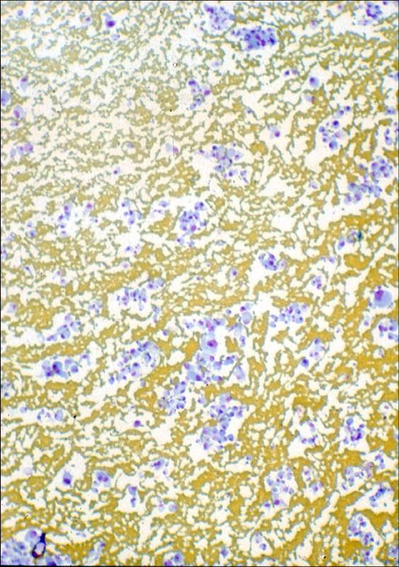
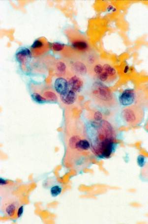
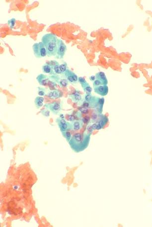
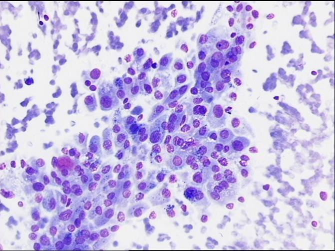
A low power view of a FNA from a Hürthle cell neoplasm (MGG). A group of rather pleomorphic Hürthle cell (Pap). A follicular group of Hürthle cells (Pap).
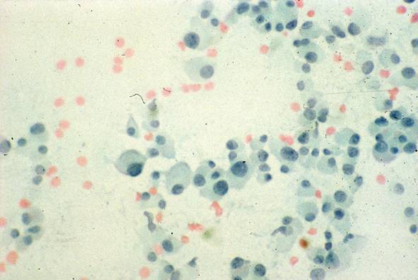
Discohesive Hürthle cells (Pap). A rather pleomorphic collection of Hürthle cells (MGG).