Lymphoma
Hodgkin and non-Hodgkin lymphomas occur as primary tumors of the lung, but spread from an extrapulmonary primary is more common. The most common primary lymphoma of the lung is low-grade extranodal marginal zone lymphoma (MALT lymphoma), followed by diffuse large B-cell lymphoma. Low grade lymphomas are typically incidental, solitary lung masses with an indolent clinical course.
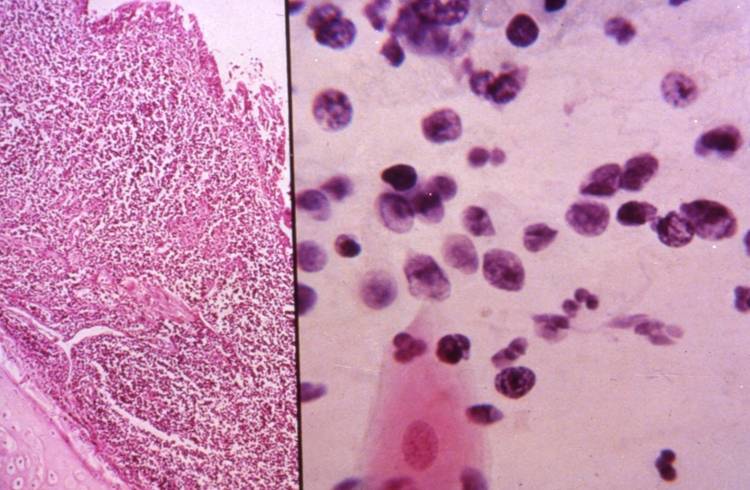
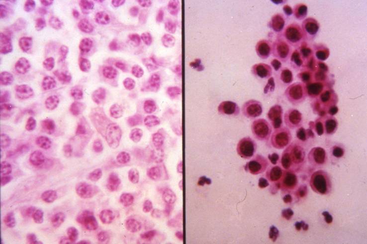
A low grade lymphoma can be particularly hard to diagnose by morphologic evaluation alone because it can be indistinguishable from an inflammatory condition. Large cell lymphomas usually show great atypia.
Differential diagnosis (non-Hodgkin lymphoma)
- Inflammatory lesions
- Small cell carcinoma
- Large cell undifferentiated carcinoma
- Melanoma
The diagnosis is facilitated when tissue fragments are available for cell block preparations. A surface marker analysis can be made to demonstrate monoclonality in low grade lymphomas. High grade lymphomas, which are obviously citologically malignant, need to stain for lymphoid markers.
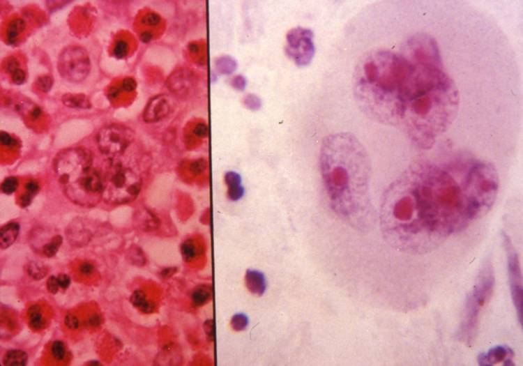
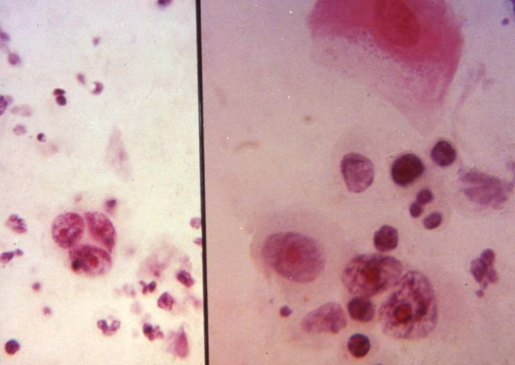
Primary Hodgkin lymphoma of the lung is very rare, while a secondary involvement is common. The cytologic diagnosis rests on identification of Reed-Sternberg cells in a background of lymphocytes, plasmacells, histiocytes and eosinophils.
Differential diagnosis (Hodgkin lymphoma)
- Granulomatous inflammatory processes
- Reactive lymphocytes (such as in ABV infections)
- Large cell non-Hodgkin lymphoma
- Melanoma
- Large cell undifferentiated carcinoma