Viral infections
Herpes simplex thacheobronchitis and pneumonitis usually affect immunocompromised patients. Multinucleated cells are commonly seen, usually with eosinophilic, intranuclear inclusions. The dignosis can be confirmed by immunoperoxidase studies.
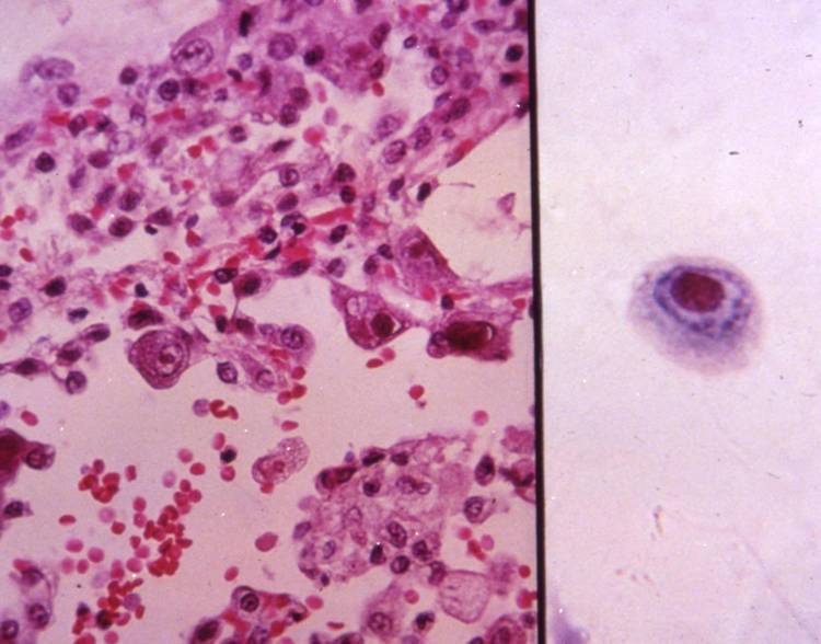
Cytomegalovirus often causes an opportunistic infection, with diffuse pulmonary infiltrates. Viral changes, which are the same as in other sites, are seen in bronchial cells or pneumocytes: the cells are enlarged, with large intranuclear inclusions with halo and small basophilic cytoplasmic inclusions. The diagnosis can be confirmed by immunoperoxidase studies, immunofluorescence or in-situ hybridization.
Measles pneumonitis usually occurs in immunocompromised children, producing a giant cell pneumonia; giant multinucleated epithelial cells, with eosinophilic intranuclear and cytoplasmic inclusions, are observed. The same findings are typical of respiratory syncitial virus infections.
In Adenovirus infections, the cells have a smudged appearance due to large inclusion filling the nucleus, and ciliocytophtoria may be pronounced: it resembles a tuft of hair and represents detachment of the terminal bar and cilia from a bronchial cell.