Mixed tumor
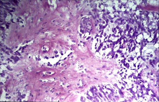
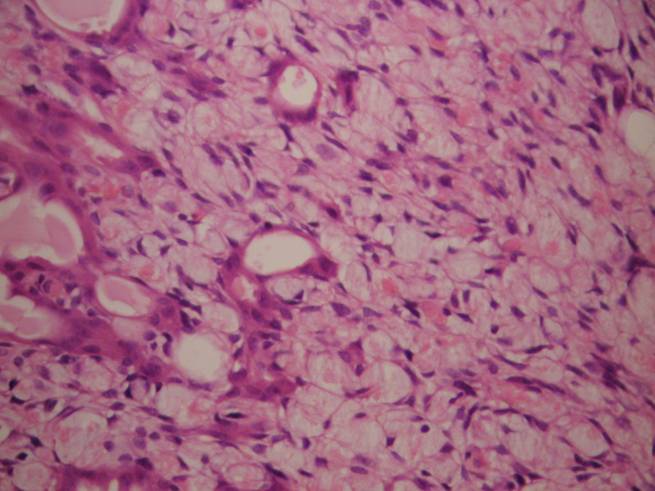
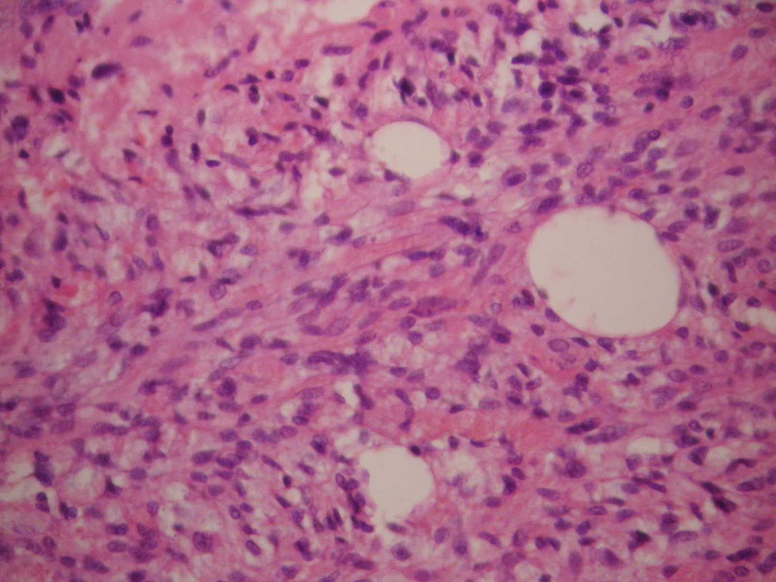
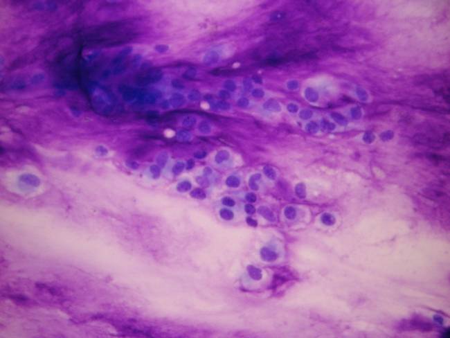
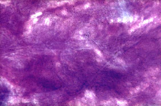
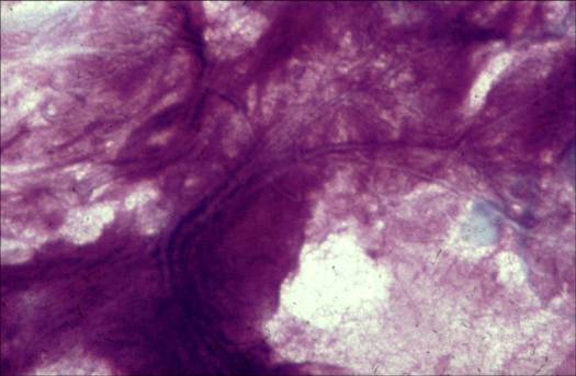
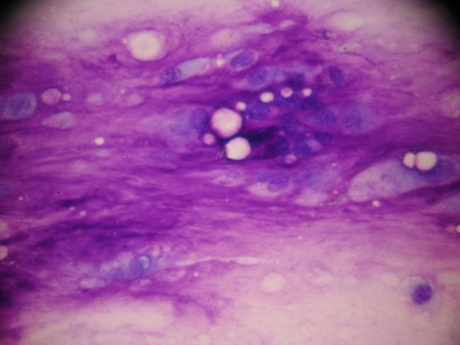
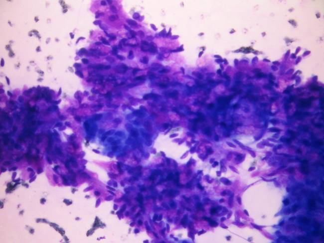
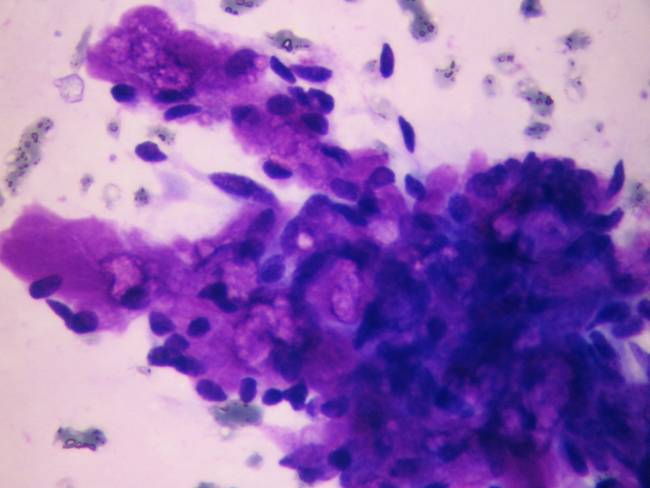
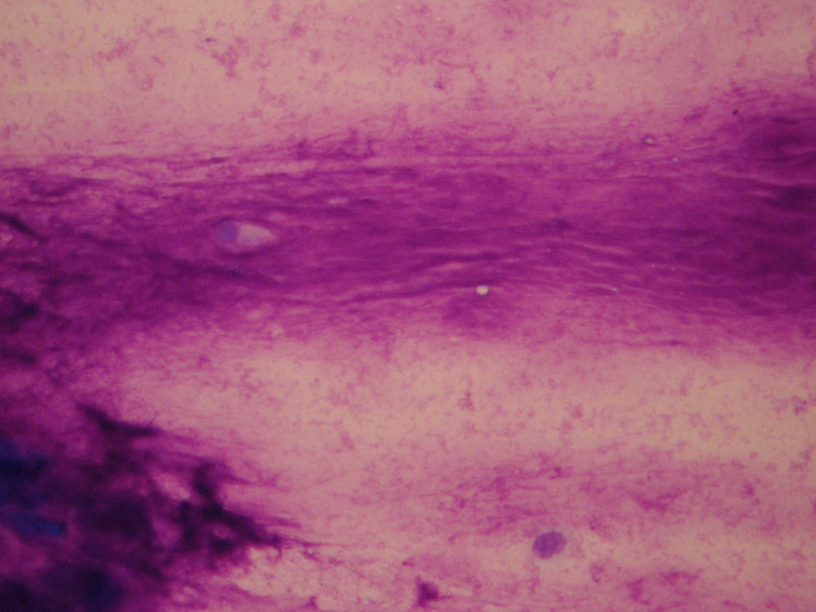
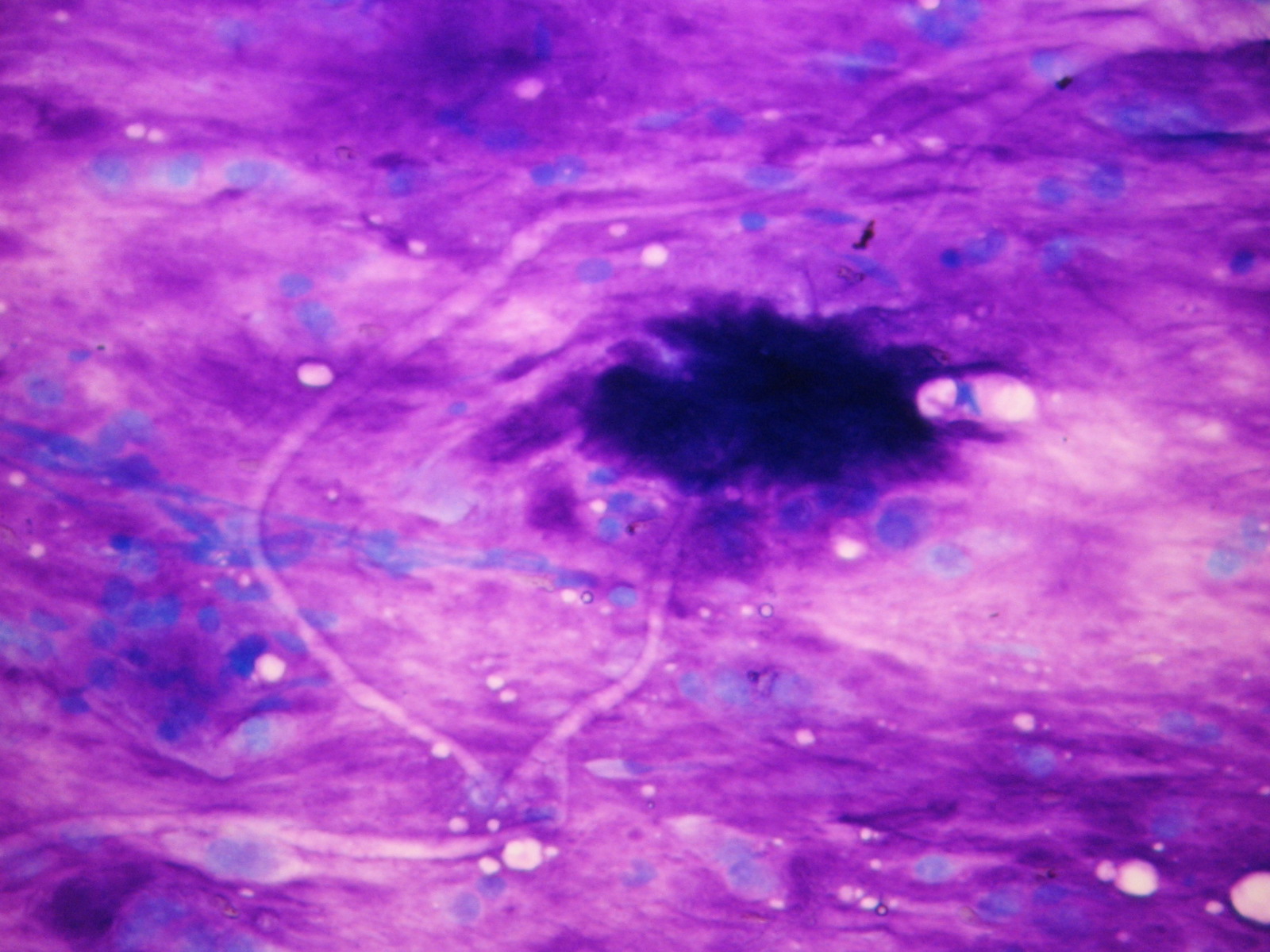
This tumor was called pleomorphic adenoma for its composite structure. It consists of myoepithelial elements, in some cases differentiating into squamous keratinizing cells or glandular structures, together with a stromal component. The myoepithelial cells may be plasmacytoid or epithelioid. Formation of cysts may occur. The stroma may be chrondroid, hyalinized, fibrotic or even osseous, usually very acellular. It is the most common tumor of the salivary glands comprising 60 – 70% of all salivary gland tumors, and arising more often in women. The complex structure of the tumour gives rise to the complex, sometimes polymorph cytologic picture. Using the Giemsa stain the chondromyxoid stromal component is metachromatic. It shows the presence of thin, pale strands in an irregularly organized pattern. This kind of stroma is diagnostic. It is not seen in adenoid cystic carcinoma.
8 – 9% of mixed tumors may undergo malignant transformation after 15 - 25 years, the result is an invasive, highly malignant tumor, usually a squamous cell or undifferentiated carcinoma. In very rare occasions spindle cell sarcomatous tumors or even carcinosarcoma can be found. The cytological malignancy of these tumors is usually simple to recognize.
Mixed tumor