Anatomy of the salivary glands
There are three pairs of main salivary glands: sublingual, submandibular and parotid gland.
The sublingual gland is the smallest in size, characterized mainly by mucinous glands. The submandibular gland is more lobulated with mixed glandular structure: there are both serous and mucinous structures in it. The parotid gland is characterized by serous acini. All of these glands have a ductal system, nerves, vessels, connective tissue and, especially the parotid gland, intraglandular lymph nodes. The number of the latter in the parotid is 30 – 50. There are several thousands of additional 'small' salivary gland all around the oral mucosal surface.Pathology (and cytopathology) of the salivary glands is very similar to that of the lachrymal glands, the sweat glands of the skin, the small submucosal glands of the trachea and bronchi.
Structure of salivary glands
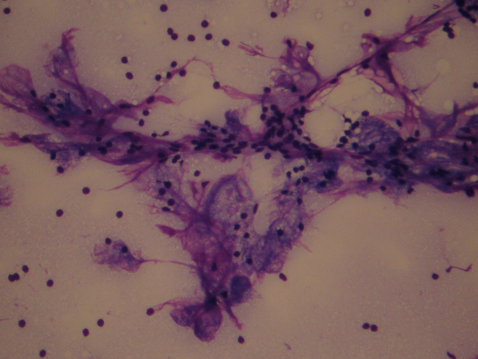
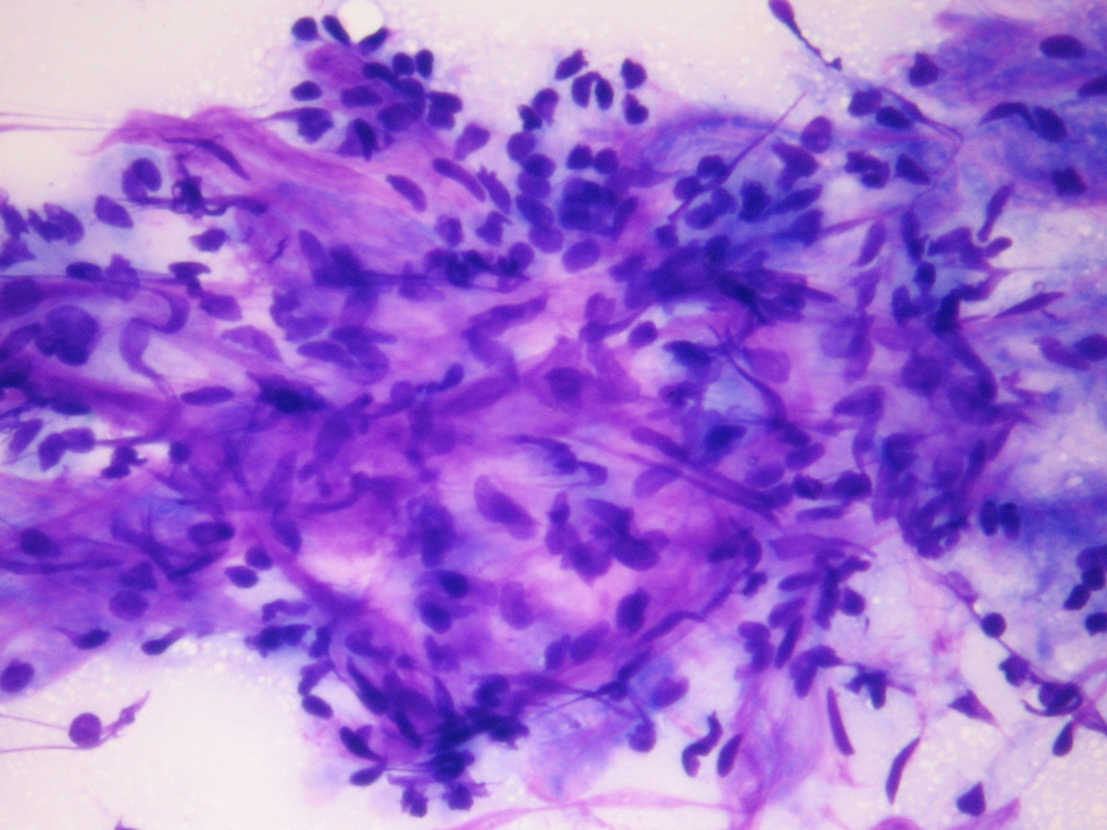
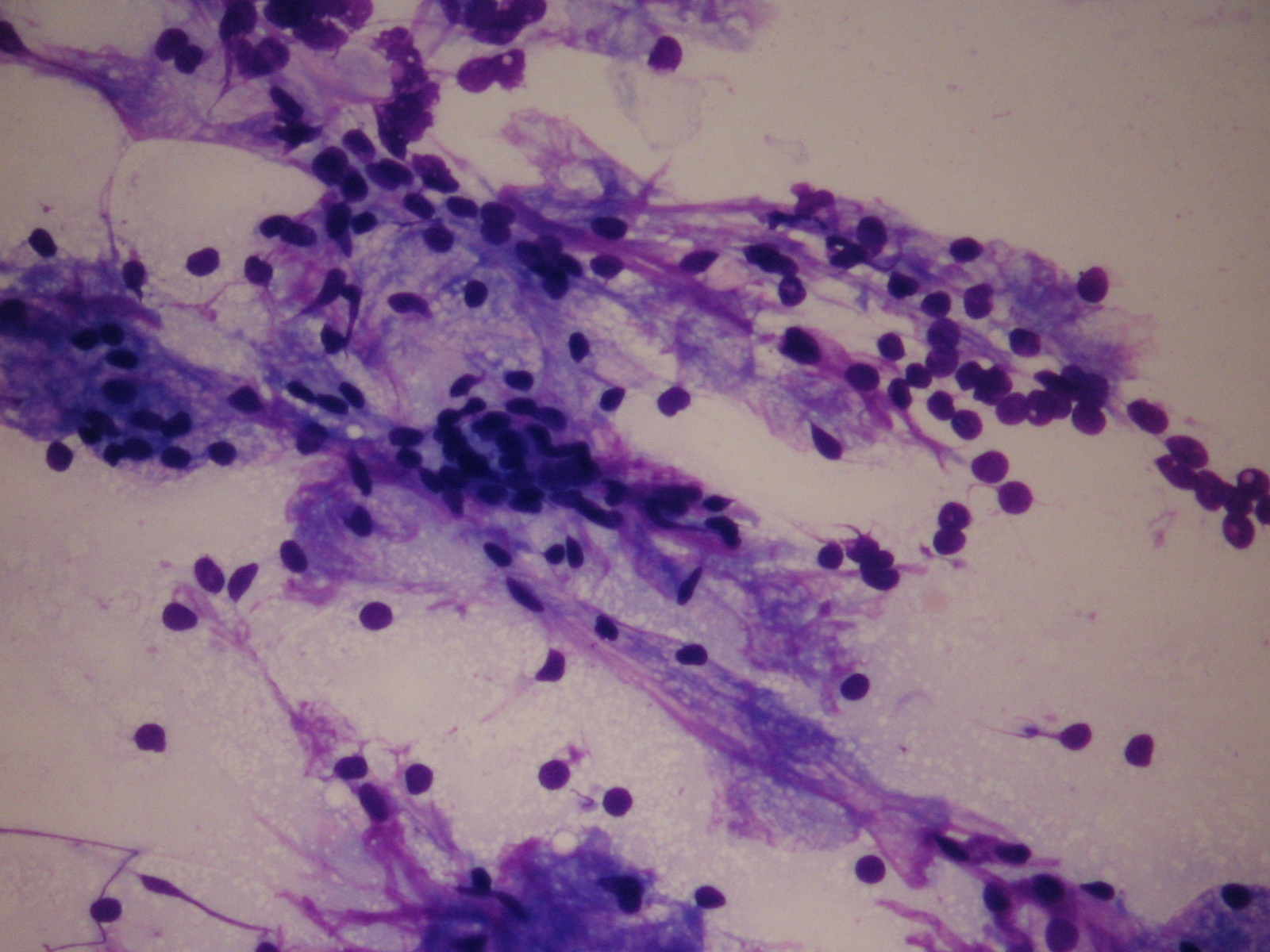
Normal cells
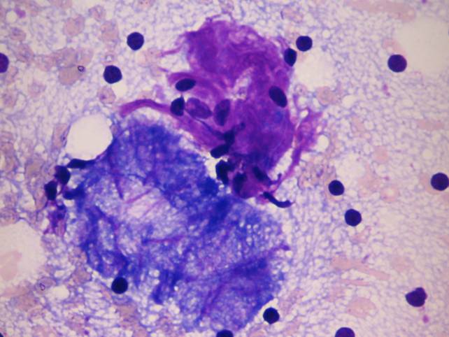
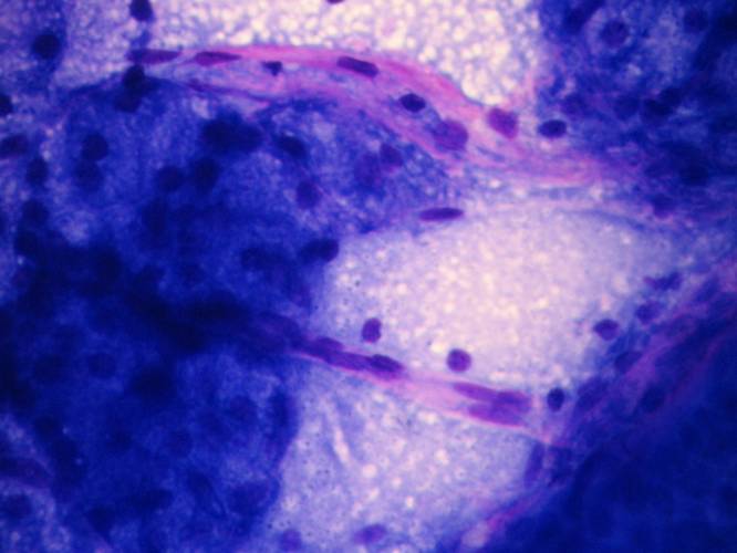
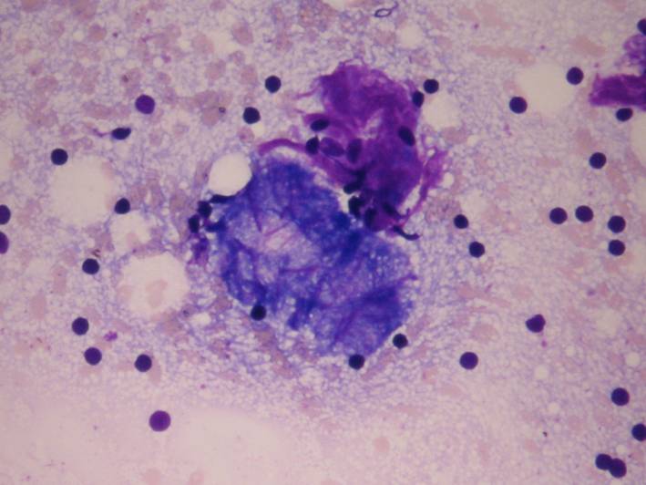
Basal cells: small cells with round nucleus. The cytoplasm is almost always euchromatic. We may find them in true tissue fragments or in three-dimensional cell clusters showing sometimes the palisading arrangement of the most 'outer' cell lines. Sometimes the background stroma is important in differentiating them from small cell tumors like adenoid cystic carcinoma: the stroma in these cases is usually metachromatic with Giemsa stain. Also the morphology of the extracellular material (cylindrical) is important.
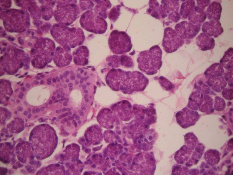
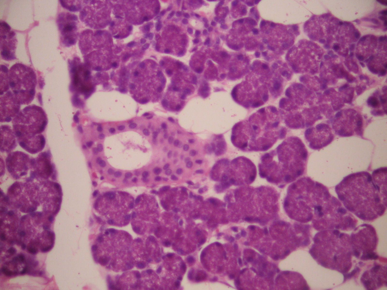
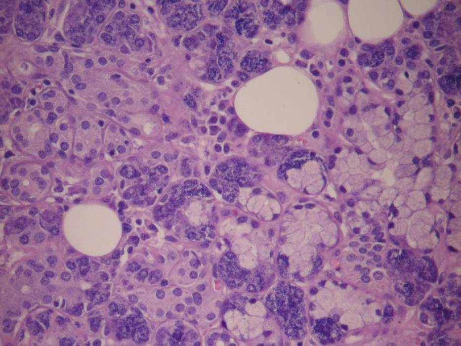
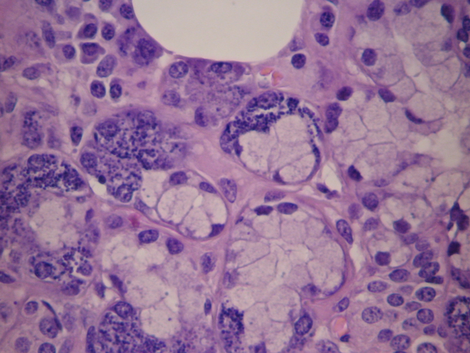
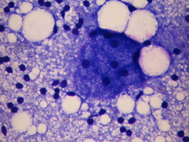
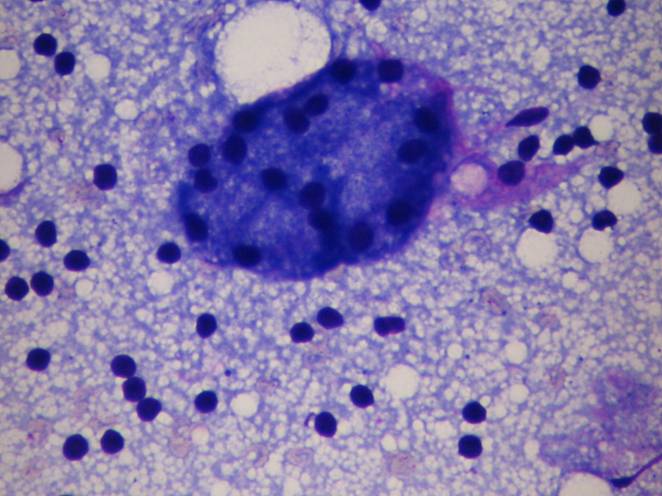
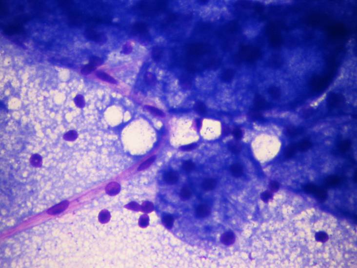
Acinar cells: large cells with serous or mucinous cytoplasmic content and small nuclei. Zymogen granules are often seen in the cytoplasm. It is very important to find ductal cells related to the acinic ones because if the former are missing we have to think of a well differentiated acinic cell carcinoma.
Ductal cells: they are usually in small honeycombed structures, the nucleus is centrally located, the cytoplasm is homogeneous. Myoepithelial cells may be attached to ductal cell fragments.
Myoepithelial cells: they are usually aspirated together with the acinic cells. They may be plasmacytoid or epithelioid with elongated cytoplasm. The nucleus is oval to round, the cytoplasm is blue and metachromatic. There are many salivary gland lesions dominated by abnormal myoepithelial cells.
Connective tissue fragments and crystalloids may also be seen.