|
Cytological diagnosis of invasive endocervical adenocarcinoma
-
While the Pap test is a sensitive test for CIN and early invasive squamous carcinoma of the cervix it is less sensitive for the detection of adenocarcinoma. The chances of detection is improved if an endocervical brush is used routinely to ensure the endocervical canal is sampled . The brush must always be used in conjunction with the spatula to ensure the transformation zone is sampled.
-
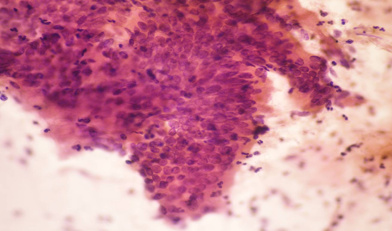
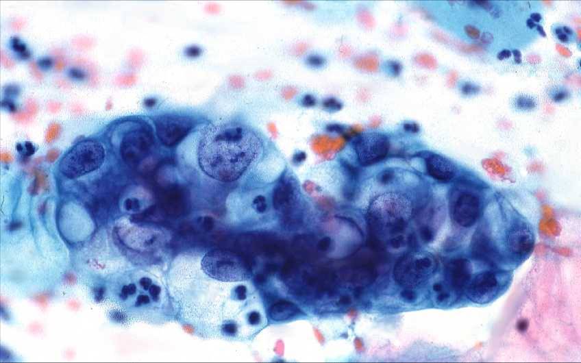
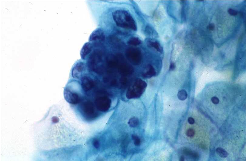
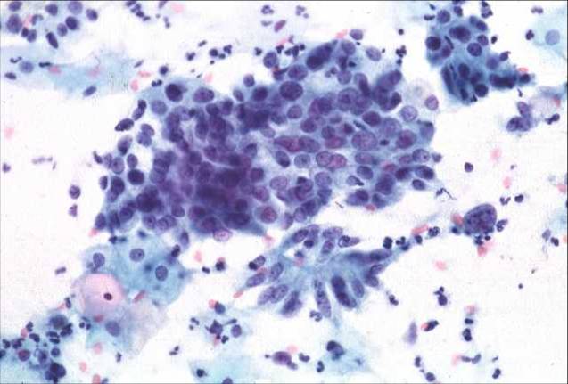
Adenocarcinoma in situ (AIS) It is difficult to distinguish between in situ adenocarcinoma and well differentiated invasive adenocarcinoma on the basis of the cytological findings. in AIS abnormal and normal endocervical epithelium may be found in close association in the same smear. In AIS is usually clean.
-
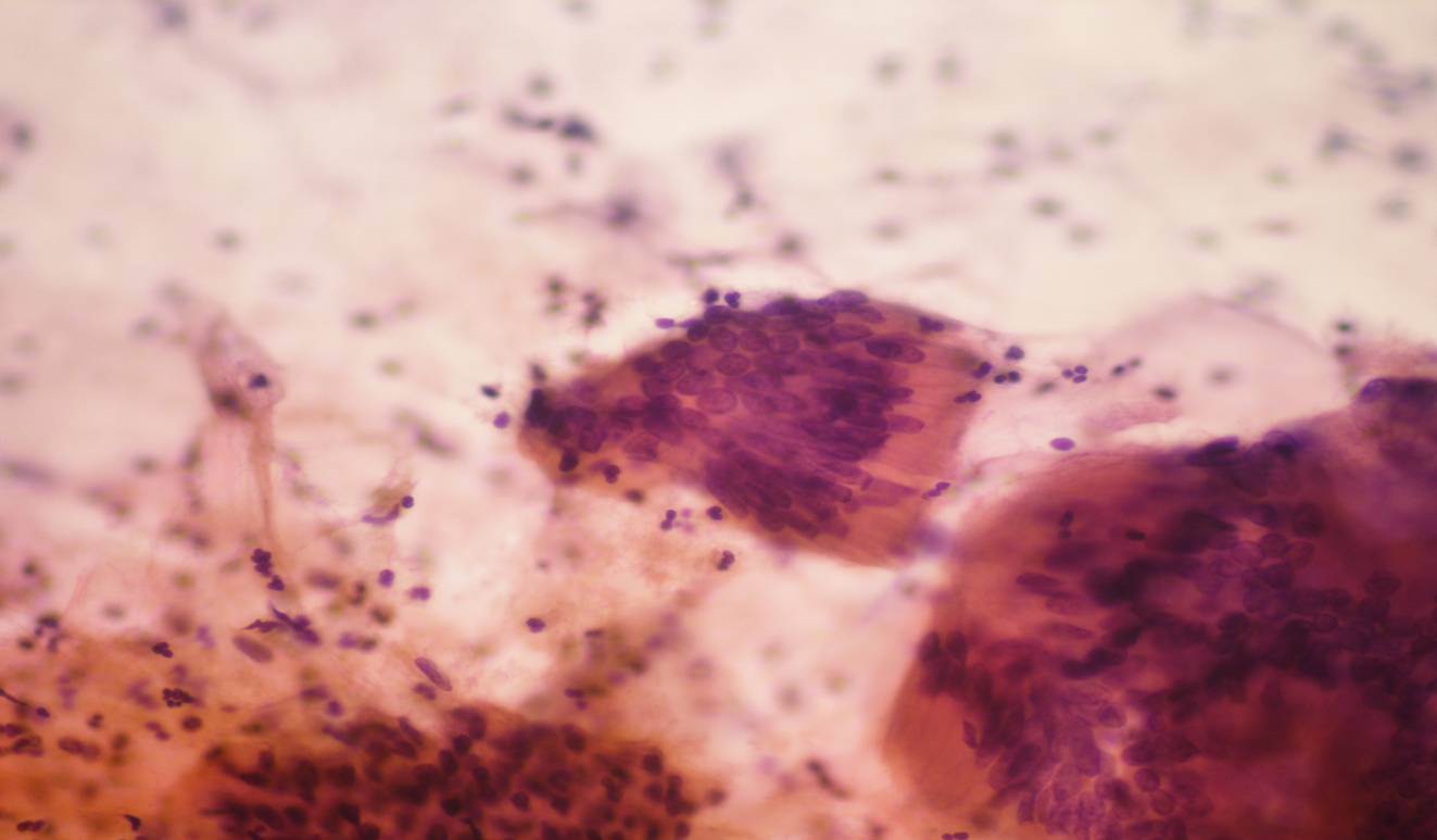
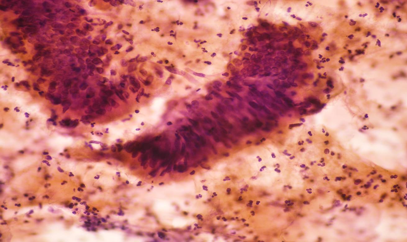
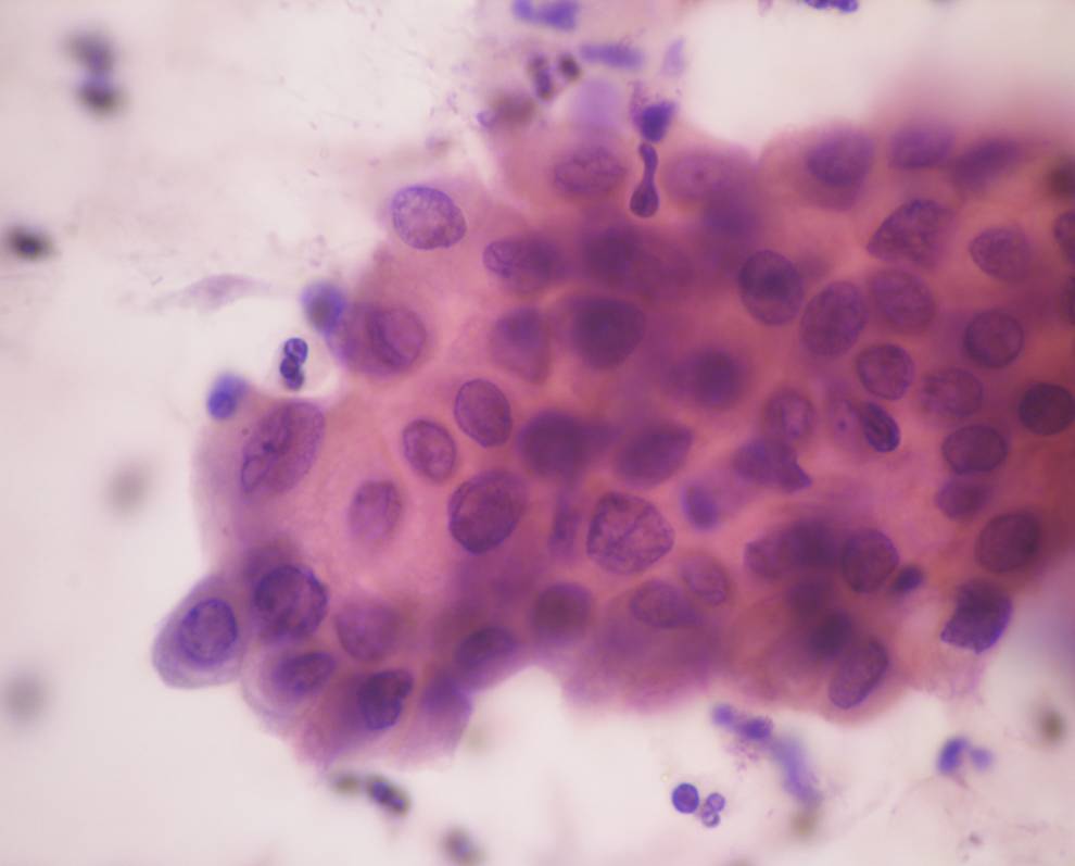
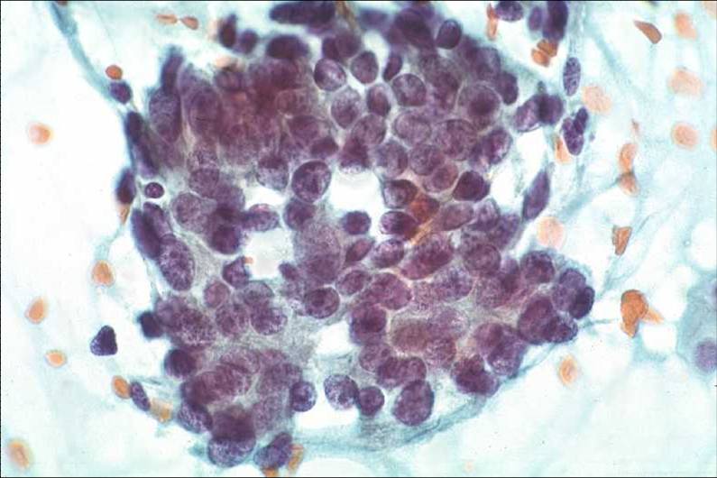
In well differentiated adenocarcinoma the glandular cells occur in sheets, clusters strips and rosettes . The individual cells have an obvious columnar appearance and may be in palisade or honey comb formation and are generally well preserved.
-
Unlike the normal palisade, the nuclei vary in size and shape. This is most apparent at the edge of the cluster.
-
Within the honey comb the nuclei are crowded and overlapping giving a hyperchromatic appearance to the group of cells although individually the nuclear chromatin may be finely granular Mitoses and nucleoli may be seen.
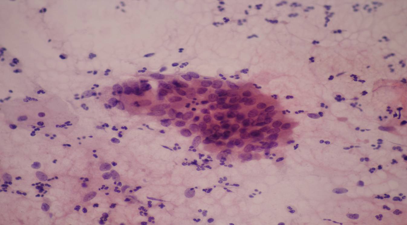
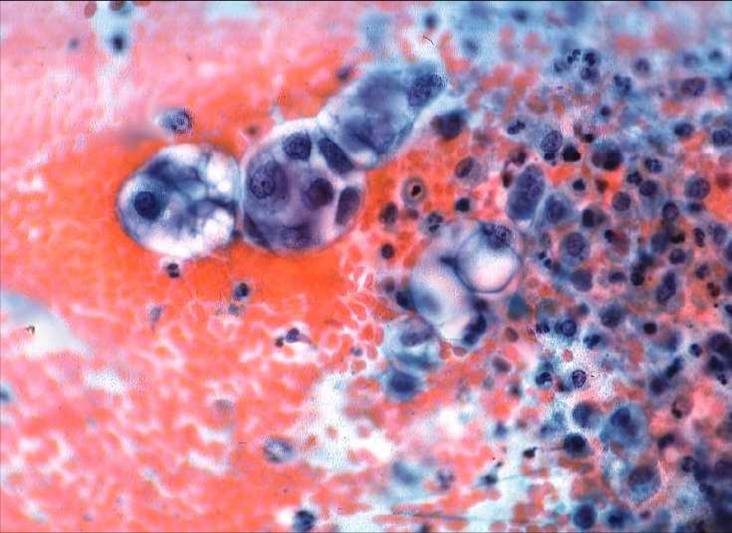
- In moderately and poorly differentiated adenocarcinomas the glandular origin of the tumour is less apparent and the cytological presentation much more variable. The cell clusters may be so densely packed that it is only by examining the edge of the tumour or the occasional discrete tumour cell that the glandular nature can be ascertained. Variation in nuclear size, enlarged and multiple nucleoli, and mitoses may be seen.
- Background : In adenocarcinoma in situ and in early stage adenocarcinoma the background to the smear is generally clean. In more advanced tumours the background may contain fresh and altered blood, polymorphs,and necrotic cell debris (the so called tumour diathesis).
- Abnormal squamous cells may be present if there is a coexisting squamous lesion.
 |
 |
Adenocarcinoma in situ |
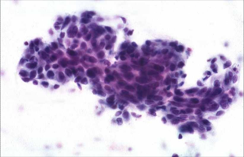 |
Poorly differentiated endocervical adenocarcinoma |
|