Anatomy, physiology and histology of the cervix
Anatomy
Endocervical canal
Ectocervix
Squamocolumnar junction, metaplastic process and Transformation zone


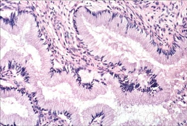
The columnar epithelium of the endocervical canal

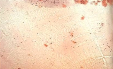
Histological section of endocervical canal Ferning of cervical mucus
  • The luminal surface of the endocervical canal and crypts is lined with a single layer of columnar cells. The lining  is made up of tall cylindrical cells arranged in a “picket “formation. The nuclei of the columnar cells are usually located in the basal part of the cell but when the cells are actively secreting mucous the nucleus may be suprabasal or in the middle of the cell. Three types of cells can be identified : secretory columnar cells, ciliated columnar cells and reserve cells..
  • The secretory cells produce  both acid and neutral mucin although the amounts vary with the menstrual cycle. The mucous is secreted by both apocrine and merocrine methods . The cervical mucous is a watery fluid whose composition varies with the stage of the menstrual cycle. It contains electrolytes especially sodium chloride and simple sugars in colloidal solution. It also contains albumins, immunoglobulins and enzymes. Leucocytes and desquamated epithelial cells are often found suspended in the mucous. The cervical mucous plays an important role in sperm transport. Mid cycle about the time of ovulation, the mucous is composed of a fine miscellar network of filaments for easy passage of the spermatozoa. At this stage “ferning “ can be demonstrated if the mucous is spread on a glass slide.  In the luteal phase of the menstrual cycle (and with oral contraceptives) the mucous becomes much thicker impeding the progress of the spermatozoa. The thickened mucous sometimes gets trapped in the endocervical crypts to form benign cysts –the so called “Nabothian follicles”.
  • The ciliated cells  are most numerous at the endometrial/endocervical junction. Their main  function is to carry the mucus along the mucous membrane
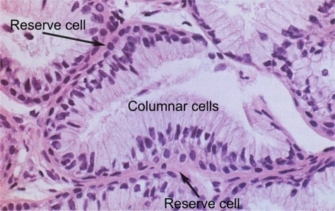
  • The reserve cells are small undifferentiated pluripotential cells lying deep to the columnar cells which are more prominent during the metaplastic process.
Columnar epithelium at higher magnification
 
Back
Next